Balbharti Maharashtra State Board 11th Biology Textbook Solutions Chapter 10 Animal Tissue Textbook Exercise Questions and Answers.
Animal Tissue Class 11 Exercise Question Answers Solutions Maharashtra Board
Class 11 Biology Chapter 10 Exercise Solutions Maharashtra Board
Biology Class 11 Chapter 10 Exercise Solutions
1. Choose correct option
Question (A)
The study of structure and arrangement of tissue is called as _______ .
(a) anatomy
(b) histology
(c) microbiology
(d) morphology
Answer:
(b) histology
![]()
Question (B)
_______ is a gland which is both exocrine and endocrine.
(a) Sebaceous
(b) Mammary
(c) Pancreas
(d) Pituitary
Answer:
(c) Pancreas
Question (C)
_______ cell junction is mediated by integrin.
(a) Gap
(b) Hemidesmosomes
(c) Desmosomes
(d) Adherens
Answer:
(b) Hemidesmosomes
Question (D)
The protein found in cartilage is _______ .
(a) ossein
(b) haemoglobin
(c) chondrin
(d) renin
Answer:
(c) chondrin
Question (E)
Find the odd one out.
(a) Thyroid gland
(b) Pituitary gland
(c) Adrenal gland
(d) Salivary gland
Answer:
(d) Salivary gland
2. Answer the following questions
Question (A)
Identify and name the type of tissues in the following:
- Inner lining of the intestine
- Heart wall
- Skin
- Nerve cord
- Inner lining of the buccal cavity
Answer:
- Epithelial tissue (Columnar epithelium)
- Cardiac muscles (Muscular tissue)
- Epithelial tissue (Stratified epithelium)
- Nervous tissue
- Epithelial tissue (Ciliated epithelium)
![]()
Question (B)
Why do animals in cold regions have a layer of fat below their skin?
Answer:
1. In adipose tissues, fats are stored in the form of droplets.
2. The adipose tissue acts as good insulator and helps retain heat in the body. This helps in survival of animals in the colder regions. Hence, animals in cold regions have a layer of fat below their skin.
Question (C)
What enables the ear pinna to be folded and twisted while the nose tip can’t be twisted?
Answer:
1. The ear pinna (outer ear) is made up of a thin plate of elastic cartilage and is connected to the surrounding.
2. The nose tip is made up of elastic cartilage. However, several bones and cartilage make up the bony- cartilaginous framework of the nose.
Hence, even though the tip of the nose is made up of elastic cartilage, it cannot be twisted like the ear pinna due to presence of bony-cartilaginous framework.
Question (D)
Sharad touched a hot plate by mistake and took away his hand quickly. Can you recognize the tissue and its type responsible for it?
Answer:
1. Nervous and muscular tissues are responsible for this action
2. Nervous tissue recognizes the stimuli whereas muscular tissue allows responding to the stimuli.
Question (E)
Priya got injured in an accident and hurt her long bone and later on she was also diagnosed with anaemia. What could be the probable reason?
Answer:
1. The centre of long bones (diaphysis) contains bone marrow, which is a site of production of blood cells (red blood cells).
2. Any severe injury to the bone marrow can affect rate of haematopoiesis (formation of blood cells).
3. A low count of erythrocytes (red blood cells) is characterised as anaemia. Hence, an injury to Priya’s long bone might have resulted in anaemia.
Question (F)
Supriya stepped out into the bright street from a cinema theatre. In response, her eye pupil shrunk. Identify the muscle responsible for the same.
Answer:
Smooth muscles (Involuntary muscles) are responsible for shrinking of eye pupil.
3. Answer the following questions
Question (A)
What is cell junction? Describe different types of cell junctions.
Answer:
1. Cell junctions: The epithelial cells are connected to each other laterally as well as to the basement
membrane by junctional complexes called cell junctions.
2. The different types of cell junctions are as follows:
a. Gap Junctions (GJs): These are intercellular connections that allow the passage of ions and small molecules between cells as well as exchange of chemical messages between cells.
b. Adherens Junctions (AJs): They are involved in various signalling pathways and transcriptional regulations.
c. Desmosomes (Ds): They provide mechanical strength to epithelial tissue, cardiac muscles and meninges.
d. Hemidesmosomes (HDs): They allow the cells to strongly adhere to the underlying basement membrane. These junctions help maintain tissue homeostasis by signalling.
e. Tight junctions (TJs): These junctions maintain cell polarity, prevent lateral diffusion of proteins and ions.
![]()
Question (B)
Describe in brief about areolar connective tissue with the help of suitable diagram.
Answer:
Areolar tissue is a loose connective tissue found under the skin, between muscles, bones, around organs, blood vessels and peritoneum. It is composed of fibres and cells.
The matrix of areolar tissues contains two types of fibres i.e. white fibres and yellow fibres.
a. White fibres: They are made up of collagen and give tensile strength to the tissue.
b. Yellow fibres: They are made up of elastin and are elastic in nature.
The four different types of cells present in this tissue are as follows:
a. Fibroblast: Large flat cells having branching processes. They produce fibres as well as polysaccharides that form the ground substance or matrix of the tissue.
b. Mast cells: Oval cells that secrete heparin and histamine.
c. Macrophages: Amoeboid, phagocytic cells.
d. Adipocytes (Fat cells): These cells store fat and have eccentric nucleus.
Question (C)
Describe the structure of multipolar neuron.
Answer:
A neuron is the structural and functional unit of the nervous tissue. A neuron is made up of cyton or cell body and cytoplasmic extensions or processes.
1. Cyton:
The cyton or cell body contains granular cytoplasm called neuroplasm and a centrally placed nucleus. The neuroplasm contains mitochondria, Golgi apparatus, RER and Nissl’s granules.
2. Cytoplasmic extensions or processes:
(a) Dendron: They are short, unbranched processes.
The fine branches of a dendron are called dendrites.
Dendrites carry an impulse towards the cyton.
(b) Axon: It is a single, elongated and cylindrical process.
- The axon is bound by the axolemma.
- The protoplasm or axoplasm contains large number of mitochondria and neurofibrils.
- The axon is enclosed in a fatty sheath called the myelin sheath and the outer covering of the myelin sheath is the neurilemma. Both the myelin sheath and the neurilemma are parts of the Schwann cell.
- The myelin sheath is absent at intervals along the axon at the Node of Ranvier.
- The fine branching structure at the end of the axon (terminal arborization) is called telodendron.
![]()
Question (D)
How to differentiate the skeletal and the smooth muscles based on their nucleus?
Answer:
Skeletal muscles contain nucleus arranged at periphery. Striated or smooth muscles are with centrally placed single large oval nucleus therefore, skeletal and smooth muscle fibres can be identified.
Question 4.
Complete the following table.
Answer:
| Cell / Tissue / Muscles | Functions |
| 1. Cardiac muscles | Cardiac muscles bring about contraction and relaxation of heart |
| 2. Tendons | Connect skeletal muscles to bones |
| 3. Chondroblast cells | Produce and maintain cartilage matrix |
| 4. Mast cells | Secrete heparin and histamine |
Question 5.
Match the following.
| ‘A’ Group | B’ Group |
| 1. Muscle | (a) Perichondrium |
| 2. Bone | (b) Sarcolemma |
| 3. Nerve cell | (c) Periosteum |
| 4. Cartilage | (d) Neurilemma |
Answer:
| ‘A’ Group | B’ Group |
| 1. Muscle | (c) Periosteum |
| 2. Bone | (a) Perichondrium |
| 3. Nerve cell | (b) Sarcolemma |
| 4. Cartilage | (d) Neurilemma |
![]()
Practical / Project:
Question 1.
To study the different tissues with the help of permanent slides in your college laboratory.
Answer:
Students may observe permanent slides of different tissues like epithelial tissue, connective tissue, muscular tissue and nervous tissue slides in laboratory.
[Students are expected to perform this activity on their own.]
Question 2.
Collect the information about the exercise to keep muscles healthy and strong.
Answer:
- Muscles become stronger when we are physically active.
- Physical activities like walking, jogging, lifting weights, playing tennis, climbing stairs, jumping, and dancing are good ways to exercise our muscles.
- Apart from this, swimming and biking can also be considered as good workouts for muscles.
- Different kinds of activities, work different muscles. Hence, it is essential to perform various types of physical activities.
- Also, activities that increase our breath rate, help in exercising our heart muscle as well.
[Students are expected to collect more information on their own.]
11th Biology Digest Chapter 10 Animal Tissue Intext Questions and Answers
Can you recall? (Textbook Page No. 116)
What is tissue?
Answer:
A group of cells having the same origin, same structure and same function is called ‘tissue’.
![]()
Do you know? (Textbook Page No. 116)
Number of cells in human body.
Answer:
There are about 100 trillion of 200 different types of cells in the human body.
Can you tell? (Textbook Page No. 119)
Explain basic structure of epithelial tissue and mention its types.
Answer:
The characteristics of epithelial tissues are as follows:
- Epithelial tissue forms a covering on inner and outer surface of body and organs.
- The cells of this tissue are compactly arranged with little intercellular matrix.
- The cells rest on a non-cellular basement membrane.
- The epithelial cells are polygonal, cuboidal or columnar in shape.
- A single nucleus is present at the centre or at the base of the cell.
- The tissue is avascular and has a good regeneration capacity.
- The major function of the epithelial tissue is protection. It also helps in absorption, transport, filtration and secretion.
The different types of epithelial tissues are as follows:
1. Simple epithelium: Epithelial tissue made up of single layer of cells is known as simple epithelium. Simple epithelium is further classified into:
a. Squamous Epithelium
b. Cuboidal Epithelium
c. Columnar Epithelium
d. Ciliated Epithelium
e. Glandular Epithelium
f. Sensory epithelium
g- Germinal epithelium
2. Compound epithelium: Epithelium composed of several layers is called compound epithelium. Compound epithelium is further classified into:
a. Stratified epithelium
b. Transitional epithelium
Epithelial tissue has good capacity of regeneration. Give reason.
Answer:
Epithelial tissue rests on a basement membrane which acts as a scaffolding on which epithelium can grow and regenerate after injuries.
![]()
Can you recall? (Textbook Page No. 116)
Where is squamous epithelium located?
Answer:
Location: It is present in blood vessels, alveoli, coelom, etc.
Can you tell? (Textbook Page No. 119)
Write a note on glandular epithelium.
Answer:
Structure:
1. The cells of the glandular epithelium can be columnar, cuboidal or pyramidal in shape.
2. The nucleus of these cells is large and situated towards the base.
3. Secretory granules are present in the cell cytoplasm.
4. Glands consist of glandular epithelium. The glands may be either unicellular (goblet cells of intestine) or multicellular (salivary gland), depending on the number of cells.
5. Types: Depending on the mode of secretion, multicellular glands can be further classified as duct bearing glands (exocrine glands) ad ductless glands (endocrine glands).
a. Exocrine glands: These glands pour their secretions at a specific site. e.g. salivary gland, sweat gland, etc.
b. Endocrine glands: These glands release their secretions directly into the blood stream, e.g. thyroid gland, pituitary gland, etc.
6. Function: Glandular epithelium secretes mucus to trap the dust particles, lubricate the inner surface of respiratory and digestive tracts, secrete enzymes and hormones, etc.
Heterocrine glands
1. Heterocrine glands or composite glands have both exocrine and endocrine function.
2. Pancreas is called a heterocrine gland because it secretes the hormone insulin into blood which is an endocrine function and enzymes into digestive tract which is an exocrine function.
![]()
Use your brain power? (Textbook Page No. 118)
When do the transitional cells change their shape?
Answer:
Transitional cells change their shape depending on the degree of distention (stretch) needed. As the tissue stretches, the transitional cells start changing shape from round and globular to thin and flat.
Can you tell? (Textbook Page No. 119)
How do cell junctions help in functioning of epithelial tissue?
Answer:
1. Cell junctions: The epithelial cells are connected to each other laterally as well as to the basement
membrane by junctional complexes called cell junctions.
2. The different types of cell junctions are as follows:
a. Gap Junctions (GJs): These are intercellular connections that allow the passage of ions and small molecules between cells as well as exchange of chemical messages between cells.
b. Adherens Junctions (AJs): They are involved in various signalling pathways and transcriptional regulations.
c. Desmosomes (Ds): They provide mechanical strength to epithelial tissue, cardiac muscles and meninges.
d. Hemidesmosomes (HDs): They allow the cells to strongly adhere to the underlying basement membrane. These junctions help maintain tissue homeostasis by signalling.
e. Tight junctions (TJs): These junctions maintain cell polarity, prevent lateral diffusion of proteins and junctions.
Can you tell? (Textbook Page No. 122)
Give reason.
As we grow old, cartilage becomes rigid.
Answer:
Calcified cartilage is a type of cartilage that becomes rigid due to deposition of salts in the matrix. This reduces the flexibility of joints in old age and cartilage becomes rigid.
Can you recall? (Textbook Page No. 116)
Enlist functions of bone.
Answer:
Bones support and protect different organs and help in movement.
![]()
Can you tell? (Textbook Page No. 122)
(i) Give reason. Bone is stronger than cartilage.
Answer:
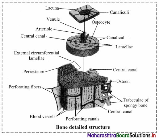
a. Bone is rigid, non-pliable, dense connective tissue characterised by the hard matrix called ossein (made up of calcium salt hydroxyapatite). An outer tough membrane called periosteum encloses the matrix. The matrix is arranged in the form of concentric layers called lamellae. Bones are well vascularized and possess blood vessels and nerves that pierce through the periosteum,
b. Cartilage is a pliable supportive connective tissue. On comparison with bones, cartilage is thin, avascular and flexible. In cartilage, a sheath of collagenous fibres called perichondrium encloses the matrix.
Hence, a bone is stronger than a cartilage.
(ii) Explain histological structure of mammalian bone.
Answer:
a. The bone is characterised by hard matrix called ossein which is made up of mineral salt hydroxy apatite (Ca10 (P04)6 (OH)2).
b. An outer tough membrane called periosteum encloses the matrix.
c. Blood vessels and nerves pierce through the periosteum.
d. The matrix is arranged in the form of concentric layers called lamellae.
e. Each lamella contains fluid filled cavities called lacunae from which fine canals called canaliculi radiate.
f. The canaliculi of adjacent lamellae connect with each other as they traverse through the matrix.
g. Active bone cells called osteoblasts and inactive bone cells called osteocytes are present in the
lacunae.
h. The mammalian bone shows the peculiar haversian system.
i. The haversian canal encloses an artery, vein and nerves.

Can you recall? (Textbook Page No. 122)
How can exercise improve your muscular system?
Answer:
1. Exercise can improve both muscular strength and stamina endurance.
2. Exercises are commonly grouped into two types depending on the effect they have on the body:
a. Aerobic exercises: such as cycling, walking, and running. They increase muscular endurance and cardiovascular health, etc.
b. Anaerobic exercises: such as weight training or sprinting, increase muscle strength, etc.
3. Anaerobic exercies: It comprises brief periods of physical exertion and high-intensity, strength-training activities.
Anaerobic exercise is a physical exercise intense enough to cause lactate to form.
It is used by athletes to promote strength, speed and power; and by body builders to build muscle mass.
![]()
Can you recall? (Textbook Page No. 122)
How many skeletal muscles are present in human body?
Answer:
There are over 650 named skeletal muscles in the human body.
Can you tell? (Textbook Page No. 125)
Differentiate between medullated and non-medullated fibre.
Answer:
| Medullated fibre | Non – Medullated fibre |
| 1. Medullary sheath is present around the axon hence also known as Myelinated nerve fibre. | Medullary sheath is absent hence also known as Non-myelinated nerve fibre. |
| 2. They have nodes of Ranvier at regular intervals. | They do not have nodes of Ranvier. |
| 3. Saltatory conduction takes place in medullated nerve fibres. | Saltatory conduction is not seen in non-medullated nerve fibre. |
| 4. These nerve fibres conduct the nerve impulse faster. | These nerve fibres conduct nerve impulse at slow rate. |
| 5. These fibres appear white in colour due to an insulating fatty layer (myelin sheath). | These fibres appear grey in colour due to absence of fatty layer. |
| 6. Schwann cell of this nerve fibre secrete myelin sheath. | Schwann cell of this nerve fibre does not secrete myelin sheath. |
| 7. Cranial nerves of vertebrates are medullated. | Nerves of autonomous nervous system are non- |
Internet is my friend. (Textbook Page No. 125)
Learn about transmission of impulse from one neuron to another.
Answer:
- A nerve impulse is transmitted from one neuron to another through junctions called synapses.
- A synapse is formed by the membranes of a pre-synaptic neuron and a post-synaptic neuron, which may or may not be separated by a gap called synaptic cleft.
- There are two types of synapses, namely, electrical synapses and chemical synapses.
- Electrical synapses: The membranes of pre- and post-synaptic neurons are in very close proximity.
Thus, electrical current can flow directly from one neuron into the other across these synapses.
Impulse transmission across an electrical synapse is faster. - Chemical synapse: The membranes of the pre- and post-synaptic neurons are separated by a fluid- filled space called synaptic cleft.
- Chemicals called neurotransmitters are involved in the transmission of impulses at these synapses.
- The axon terminals contain vesicles filled with these neurotransmitters.
- When an impulse arrives at the axon terminal, it stimulates the movement of the synaptic vesicles towards the membrane where they fuse with the plasma membrane and release their neurotransmitters into the synaptic cleft.
- The released neurotransmitters bind to their specific receptors, present on the post-synaptic membrane.
- This binding opens ion channels and allows the entry of ions which can generate a new potential in the post-synaptic neuron.
[Students are expected to refer the given information and collect more information from the internet.]
[Note: Students can scan the adjacent QR code to get conceptual clarity with the aid of a relevant video ]
![]()
Observe and Discuss (Textbook Page No. 125)
Explain the structure of nerve.

Answer:
- Each spinal nerve consists of many axons and contains layers of protective connective tissue coverings.
- Axons are enclosed in a fatty sheath called myelin sheath.
- Individual axons within a nerve are wrapped in an endoneurium (innermost layer).
- Groups of axons with their endoneurium are arranged in bundles called fascicles.
- Each fascicle is wrapped in perineurium (middle layer).
- The outermost covering over the entire nerve is the epineurium. The epineurium extends between fascicles.
- Many blood vessels nourish the nerve and are present within the perineurium and epineurium.
[Source: Tortora. G, Derrickson. B. Principles of Anatomy and Physiology. 11th Edition.]
11th Std Biology Questions And Answers:
- Morphology of Flowering Plants Class 9 Biology Questions And Answers
- Animal Tissue Class 9 Biology Questions And Answers
- Study of Animal Type : Cockroach Class 9 Biology Questions And Answers
- Photosynthesis Class 9 Biology Questions And Answers
- Respiration and Energy Transfer Class 9 Biology Questions And Answers
- Human Nutrition Class 9 Biology Questions And Answers
- Excretion and Osmoregulation Class 9 Biology Questions And Answers
- CSkeleton and Movement Class 9 Biology Questions And Answers
