Balbharti Maharashtra State Board 11th Biology Textbook Solutions Chapter 8 Plant Tissues and Anatomy Textbook Exercise Questions and Answers.
Plant Tissues and Anatomy Class 11 Exercise Question Answers Solutions Maharashtra Board
Class 11 Biology Chapter 8 Exercise Solutions Maharashtra Board
Biology Class 11 Chapter 8 Exercise Solutions
1. Choose the correct option.
Question (A)
Location or position of meristematic regions is divided into _______ types.
(A) one
(B) two
(C) three
(D) none of the above
Answer:
(C) three
![]()
Question (B)
Cambium is also called
(A) apical meristem
(B) intercalary meristem
(C) lateral meristem
(D) none of the above
Answer:
(C) lateral meristem
Question (C)
Collenchyma is a type of ________ tissue.
(A) living
(B) dead
(C) living and dead
(D) none of the above
Answer:
(A) living
Question (D)
_______ is a complex permanent tissue.
(A) Parenchyma
(B) Sclerenchyma
(C) Chlorenchyma
(D) Xylem
Answer:
(D) Xylem
Question (E)
Mesophyll tissue is present in ________ .
(A) root
(B) stem
(C) leaf
(D) flower
Answer:
(C) leaf
![]()
2. Answer the following questions
Question (A)
A fresh section was taken by a student but he was very disappointed because there were only few green and most colourless cells. Teacher provided a pink colour solution. The section was immersed in this solution and when observed it was much clearer. What is the magic?
Answer:
1. The pink coloured solution given by teacher must be a saffanin stain.
2. Saffanin is used to stain plant tissues, especially lignified tissues such as cell wall and xylem.
Question (B)
While observing a section, many scattered vascular bundles could be seen. Teacher said, in spite of this large number the stem cannot grow in girth. Why?
Answer:
- Students must have observed monocot stems.
- It is because, monocot stem shows scattered vascular bundles.
- In monocot stem, vascular bundles are closed i.e. without cambium.
- Thus, secondary growth does not occur which is required for increase in girth. Hence, in spite of having large number of scattered vascular bundles, monocot stems do not grow in girth.
Question (C)
A section of the stem had vascular bundles, where one tissue was wrapped around the other. How will you technically describe it?
Answer:
Concentric vascular bundle:
a. When one vascular tissue is completely encircling the other, it is called as concentric vascular bundle.
b. When phloem is encircled by xylem, it is called as leptocentric vascular bundle, whereas when xylem is encircled by phloem, it is called as hadrocentric vascular bundle.
c. When xylem is encircled by phloem on both faces, it is called as amphicribral vascular bundle. When phloem is encircled by xylem on both faces it is called as amphivasal vascular bundle.
Question (D)
There were two cut logs of wood lying in the campus. One had growth rings and other didn’t. Teacher said it is due to differences in their pattern of grow th which is dependent on season. How?
Answer:
1. It is possible that one of the cut logs was of a tropical tree, whereas the other was of a temperate tree. Since tropical trees grow in a similar manner all year, growth rings are not apparent. Another explanation for this could be that the log which had growth rings must be of an old tree which has experience many seasons, whereas the log without growth rings must be of younger tree, that has not been subjected to seasonal changes and hence not developed prominent growth rings.
2. Growth rings are formed due cambial activity during favourable and non-favourable climatic conditions.
3. During favourable conditions, spring wood (early wood) is formed which has broader xylem bands, lighter colour, tracheids with thin wall and wide lumen, fibres are less in number, low density. Whereas, during unfavourable conditions, autumn wood (late wood) is formed which has narrow xylem band, darker in colour, lumen is narrow and walls are thick with abundant fibres, high density.
4. Spring wood and autumn wood that appear as alternate light and dark concentric rings, constitute an annual ring or growth ring.
5. These growth rings can be used to estimate the age of the tree. These are found more in older trees as compare to younger tree.
Question (E)
While on the trip to Kashmir, Pintoo observed that cut portions of large trees show distinct rings, which he never found in Maharashtra. Why is so?
Answer:
1. Cut portions of large tress show distinct rings which are annual rings formed due to activity of cambium during favourable and non-favourable climatic conditions.
2. Kashmir falls under temperate region where the climatic conditions are not uniform through the year. In the spring season, conditions are favourable due to which cambium is active, whereas in autumn season, conditions are unfavourable due to which cambium is less active. This leads to formation of spring wood and autumn wood that appear as alternate light and dark concentric rings, constitute an annual ring or growth ring.
3. Maharashtra falls under tropical region where climatic conditions are favourable throughout the year. In tropical areas, continuous growth of secondary xylem occurs. Thus, trees growing in tropical regions show less or no annual rings as compared to trees in temperate region.
![]()
Question (F)
A student was observing a slide with no label under microscope. The section had some vascular bundles scattered in the ground tissue. It is section of a monocot stem! He exclaimed. No! it is section of fern rachis, said the teacher. Teacher told to observe vascular bundle again. Student agreed, Why?
Answer:
- In fern rachis, the number of vascular bundles is less as compared to number of vascular bundles in monocot stem. In monocot stem, vascular bundles are numerous.
- In fern rachis, xylem consists of only tracheids whereas in monocot stem, xylem consists of vessels (protoxylem and metaxylem) as well as tracheids. Monocot stem shows presence of lysigenous cavity just below protoxylem.
- In fern rachis, phloem consists of only sieve cells whereas in monocot stem, phloem consists of sieve tubes and companion cells. Thus, a student must hav e observed these differences in the given section and agreed to teacher’s statement that the given section is of fern rachis and not of monocot stem.
Question (G)
Student found a wooden stopper in lab. He was told by an old lab attendant that it is there for many years. He kept thinking how it did not rot?
Answer:
1. Wooden stopper or cork is obtained from the phellem (cork) part of a bark.
2. Phellem (cork) is impervious in nature and does not allow entry of water due to suberized walls.
3. Due to this it does not rot and remains as it is for many years.
Question (H)
Student while observing a slide of leaf section observed many stomata on the upper surface. He thought he has placed slide upside down. Teacher confirmed it is rightly placed. Explain.
Answer:
1. In a dicot leaf, stomata are generally absent on upper epidermis but are present on lower epidermis. Thus, the student must have thought that he has placed slide upside down.
2. According to teacher, the section was placed rightly, thus the given section must be of monocot leaf.
3. It is because, in monocot leaf stomata are present on both upper and lower epidermis.
![]()
3. Write short notes on the following points.
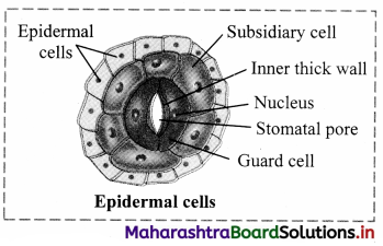
Question (A)
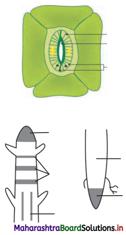
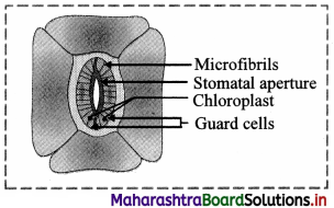
Structure of stomata.
Answer:
- Small gateways in the epidermal cells are called as stomata.
- Stoma is controlled or guarded by specially modified cells called guard cells.
- These guard cells may be kidney-shaped (dicot) or dumbbell-shaped (monocot), collectively called as stomata.
- Guard cells have chloroplasts to carry out photosynthesis.
- Change in turgor pressure of guard cells causes opening and closing of stomata, which enables exchange of gases and water vapour.
- Stomata are further covered by subsidiary cells.
- Stoma, guard cells and subsidiary cells form a unit called stomatal apparatus.
Question (B)
Write a short note on secondary growth.
OR
With the help of neat and labelled diagram explain the secondary growth in dicot stem.
Answer:
Secondary growth:
- Dicotyledonous plants and gymnosperms exhibit increase in girth of root and stem.
- In dicot stem, secondary growth begins with the formation of a continuous cambium ring.
- The cambium present between the primary xylem and primary phloem of a vascular bundle is called intrafascicular cambium.
- The cells of medullary rays adjoining these intrafascicular cambium strips become meristematic (regain the capacity to divide) and form the interfascicular cambium.
- Thus, a complete and continuous ring of vascular cambium is formed.
- The cambium ring cuts off new cells, towards both inner and outer sides.
- The cells that are cut-off towards pith (inner side) mature into secondary xylem and cells that are cut-off towards periphery mature into secondary phloem.
- Generally, amount of secondary xylem is more than the secondary phloem.
![]()
Question (C)
Write a short note on peculiarity of a sclerenchyma cell wall.
Answer:
Peculiarity of a sclerenchyma cell wall:
1. Cell wall of sclerenchyma is evenly thickened due to uniform deposition of lignin.
2. Cell wall of sclereids is extremely thick and strongly lignified.
4. Differentiate
Question (A)
Differentiate between vascular bundles of monocot and dicot.
Answer:
- Vascular bundle of monocot and dicot root.
- Vascular bundle of monocot and dicot stem.
- Vascular bundle of monocot and dicot leaf.
Question (B)
Differentiate between xylem and phloem.
Answer:
| Xylem | Phloem |
| 1. It is a dead complex tissue. | It is a living complex tissue. |
| 2. It is composed of xylem, tracheids, vessels, xylem fibres and xylem parenchyma. | It is composed of sieve tubes, sieve cells, companion cells, phloem parenchyma and phloem fibres. |
| 3. It is also known as wood. | It is also known as bast. |
| 4. The cell walls are thick due to lignin. | The cell walls are thin. |
| 5. Xylem conducts water and minerals from roots to the stem and leaves. It also provides mechanical strength to the plant parts. | It is the chief food conducting tissue of vascular plants responsible for translocation of food from leaves to other plant parts. |
![]()
5. Draw neat labelled diagrams
Question (A)
T.S. of dicot leaf.
Answer:
1. Structure of dorsiventral leaf: The mesophyll tissue is differentiated into palisade and spongy parenchyma in a dorsiventral leaf. This type is very common in dicot leaf. The different parts of this leaf are as follows:
2. Upper epidermis: It consists of a single layer of tightly packed rectangular, barrel shaped, parenchymatous cells which are devoid of chloroplast. A distinct layer of cuticle lies on the outside of the epidermis. Stomata are generally absent.
3. Mesophyll: Between upper and lower epidermis, there is chloroplast-containing photosynthetic tissue called mesophyll It is differentiated into Palisade parenchyma and Spongy parenchyma.
a. Palisade parenchyma:
Palisade parenchyma is present below upper epidermis and consists of closely packed elongated cells. The cells contain abundant chloroplasts and help in photosynthesis.
b. Spongy parenchyma:
Spongy parenchyma is present below palisade tissue and consists of loosely arranged irregularly shaped cells with intercellular spaces. The spongy parenchyma cells contain chloroplast and are in contact with the atmosphere through stomata.
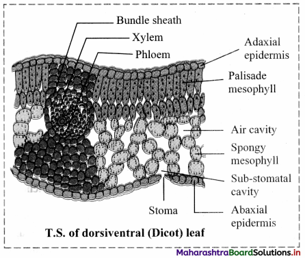
4. Vascular system: It is made up of a number of vascular bundles of varying size depending upon the venation. Each one is surrounded by a thin layer of parenchymatous cells called bundle sheath. Vascular bundles are closed. Xylem lies towards upper epidermis and phloem towards lower epidermis. Cambium is absent, hence there is no secondary growth in the leaf.
5. Lower epidermis: It consists of a single layer of compactly arranged rectangular, parenchymatous cells. A thin layer of cuticle is also present. The lower epidermis contains a large number of microscopic pores called stomata. There is an air-space called substomatal chamber at each stoma.
![]()
Question (B)
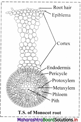
T.S. of Monocot root.
Answer:
Question (C)
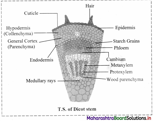
Draw neat labelled diagrams of T.S. of dicot stem.
Answer:
Question 6.
Write the information related to diagram given below.
Answer:
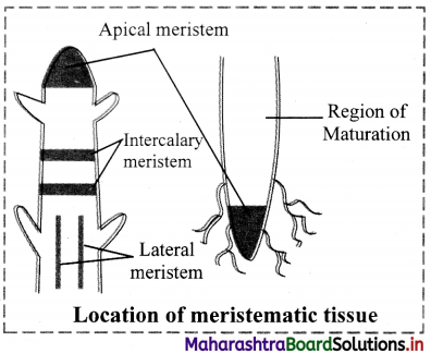
[Note: The labelled part can be considered as the ‘region of maturation ’ of root apical however, the region of maturation does not contain meristematic tissue ]
Classification of meristematic tissue based on its position:
1. Apical meristem:
a. It is produced from promeristem and forms growing point of apices of root, shoot and their lateral branches.
b. It brings about increase in length of plant body and is called as apical initials.
c. Shoot apical meristem is terminal in position whereas in root it is subterminal i.e. located behind the root cap.
2. Intercalary meristem:
a. Intercalary meristematic tissue is present in the top or base area of node.
b. Their activity is mainly seen in monocots.
c. These are short lived.
3. Lateral meristem:
a. It is present along the sides of central axis of organs.
b. It takes part in increasing girth of stem or root, e.g. Intrafascicular cambium.
c. It is found in vascular bundles of gymnosperms and dicot angiosperms.
![]()
Question 7.
Identify the following diagrams, label it and prepare a chart of characteristics.
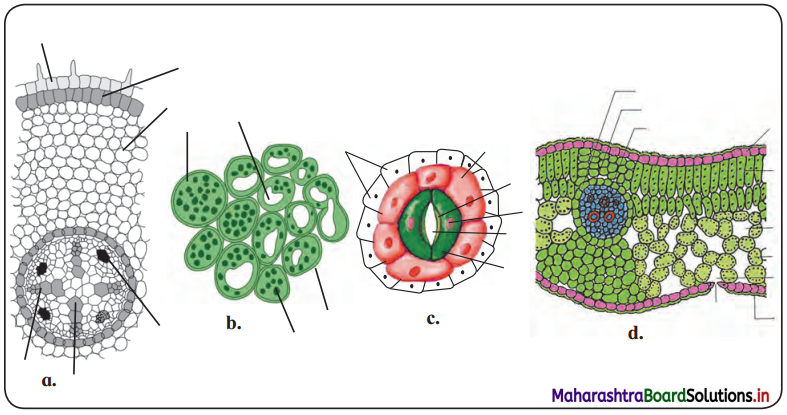 Answer:
1. Figure ‘c’
Question 8.
Distinguish between dicot and monocot leaf on the basis of following characters.
Answer:
| Characters | Dicot leaf | Monocot leaf |
| Stomata | Stomata are restricted to lower epidermis. Guard cells of stoma are kidney shaped. | Stomata occur on both epidermis. Guard cells of stoma are dumbbell shaped. |
| Intercellular space | More intercellular spaces due to presence of spongy parenchyma. | Less intercellular spaces as mesophyll is not differentiated into spongy and palisade tissue. |
| Venation | Reticulate venation | Parallel venation |
| Vascular bundle | Vascular bundles of varying size. The size of the vascular bundles is dependent on the size of the veins which vary in thickness in dicot leaf. | Vascular bundles are nearly of similar size (Except in main veins). |
| Mesophyll cells | Mesophyll tissue is differentiated into palisade parenchyma and spongy parenchyma. | Mesophyll tissue is not differentiated into palisade parenchyma and spongy parenchyma. |
![]()
Practical/ Project:
Question 1.
Prepare detail anatomical charts with diagrammatic representation of dicot and monocot plants.
Answer:
Anatomy of dicot root: The transverse section of a typical dicotyledonous root shows following anatomical features:
1. Epiblema: It is the outermost single layer of cells without cuticle. Some epidermal cells prolong to form unicellular root hairs.
2. Cortex: It is made up of many layers of thin walled parenchyma cells. Cortical cells store food and water.
3. Exodermis: After the death of epiblema, outer layer of cortex become cutinized and is called Exodermis.
4. Endodermis:
The innermost layer of cortex is called Endodermis.
The cells are barrel-shaped and their radial walls bear Casparian strip or Casparian bands composed of suberin. Near the protoxylem, there are unthickened passage cells.
5. Stele: It consists of pericycle, vascular bundles and pith.
a. Pericycle: Next to the endodermis, there is a single layer of thin walled parenchyma cells called pericycle. It forms outermost layer of stele or vascular cylinder.
b. Vascular bundle: Vascular bundles are radial. Xylem and Phloem occur in separate patches arranged on alternate radii. Xylem is exarch in root that means protoxylem vessels are towards periphery and metaxylem elements are towards centre. Xylem bundles vary from two to six number, i.e. they may be diarch, triarch, tetrarch, etc.
Connective tissue: A parenchymatous tissue is present in between xylem and phloem.
c. Pith: The central part of stele is called pith. It is narrow and made up of parenchymatous cells, with or without intercellular spaces.
6. At a later stage cambium ring develops between the xylem and phloem causing secondary growth.
Anatomy of monocot stem: A transverse section of maize (monocot) stem shows the following structures:
- Epidermis: It is single-layered and without trichomes.
- Hypodermis: It is sclerenchymatous.
- Ground tissue: It consists of thin walled parenchyma cells. It extends from hypodermis to the centre. It is not differentiated into cortex, endodermis, pericycle and pith.
- Vascular bundles: Vascular bundles are numerous and are scattered in ground tissue. Each vascular bundle is surrounded by a sclerenchymatous bundle sheath. Vascular bundles are conjoint, collateral and closed (without cambium). Xylem is endarch and shows lysigenous cavity.
- Pith: Pith is absent.
Anatomy of dicot leaf:
1. Structure of dorsiventral leaf: The mesophyll tissue is differentiated into palisade and spongy parenchyma in a dorsiventral leaf. This type is very common in dicot leaf. The different parts of this leaf are as follows:
2. Upper epidermis: It consists of a single layer of tightly packed rectangular, barrel shaped, parenchymatous cells which are devoid of chloroplast. A distinct layer of cuticle lies on the outside of the epidermis. Stomata are generally absent.
3. Mesophyll: Between upper and lower epidermis, there is chloroplast-containing photosynthetic tissue called mesophyll It is differentiated into Palisade parenchyma and Spongy parenchyma.
a. Palisade parenchyma:
Palisade parenchyma is present below upper epidermis and consists of closely packed elongated cells. The cells contain abundant chloroplasts and help in photosynthesis.
b. Spongy parenchyma:
Spongy parenchyma is present below palisade tissue and consists of loosely arranged irregularly shaped cells with intercellular spaces. The spongy parenchyma cells contain chloroplast and are in contact with the atmosphere through stomata.
4. Vascular system: It is made up of a number of vascular bundles of varying size depending upon the venation. Each one is surrounded by a thin layer of parenchymatous cells called bundle sheath. Vascular bundles are closed. Xylem lies towards upper epidermis and phloem towards lower epidermis. Cambium is absent, hence there is no secondary growth in the leaf.
5. Lower epidermis: It consists of a single layer of compactly arranged rectangular, parenchymatous cells. A thin layer of cuticle is also present. The lower epidermis contains a large number of microscopic pores called stomata. There is an air-space called substomatal chamber at each stoma.
Anatomy of monocot leaf:
1.It is single layered, present on both sides of the leaf.
It consists of compactly arranged rectangular transparent parenchymatous cells.
Both the surfaces contain stomata.
Both the surfaces have a distinct layer of cuticle.
2. Mesophyll:
Mesophyll is not differentiated into palisade and spongy tissue.
3. Vascular bundle:
These are conjoint, collateral and closed.
![]()
Question 2.
Observe different slides related to anatomy of flowering plants under the guidance of teacher.
[Students are expected to perform this practical own their own.]
11th Biology Digest Chapter 8 Plant Tissues and Anatomy Intext Questions and Answers
Can you recall? (Textbook Page No. 85)
(i) Which component brings about important processes in the living organisms?
Answer:
Cell is the component that brings about important processes in the living organisms.
(ii) What is tissue?
Answer:
A group of cells having essentially a common function and origin is called as tissue.
![]()
(iii) Explain simple and complex tissue.
Answer:
a. Simple tissue:
1. They are made up of only one type of cells.
2. They are found in all the plant parts.
3. They perform many functions.
4. Simple tissues in plants are Parenchyma, Collenchyma, Sclerenchyma.
b. Complex tissue:
1. They are made up of many types of cells.
2. They are found only in the vascular regions of the plant.
3. They mainly perform the function of conduction of food and water.
4. Complex tissues in plants are Xylem and Phloem.
(iv) Complete the flow chart.
Organisms → Organs → Cells
Answer:
Organism → Organ system → Organs → Tissue system → Tissue → Cells
Can you tell? (Textbook Page No. 86)
Enlist the characteristics of meristematic tissue.
Answer:
Characteristics of meristematic tissue:
- It is a group of young, immature cells.
- These are living cells with ability to divide in the regions where they are present.
- These are polyhedral or isodiametric in shape without intercellular spaces.
- Cell wall is thin, elastic and mainly composed of cellulose.
- Protoplasm is dense with distinct nucleus at the centre and vacuoles if present, are very small.
- Cells show high rate of metabolism.
Can you tell? (Textbook Page No. 86)
Classify meristematic tissue on the basis of origin.
Answer:
Classification of meristematic tissue on the basis of origin:
1. Promeristem / Primordial meristem:
a. It is also called as embryonic meristem.
b. It usually occupies very minute area at the tip of root and shoot.
2. Primary meristem:
a. It originates from the primordial meristem and occurs in the plant body from the beginning, at the root and shoot apices.
b. Cells are always in active state of division and give rise to permanent tissues.
3. Secondary meristem:
a. These tissues develop from living permanent tissues during later stages of plant growth hence are called as secondary meristems.
b. This tissue occurs in the mature regions of root and shoot of many plants.
c. Secondary meristem is always lateral (to the central axis) in position e.g. Fascicular cambium, inter fascicular cambium, cork cambium.
![]()
Can you tell? (Textbook Page No. 89)
Write a note on parenchyma.
Answer:
Parenchyma:
- It is a type of simple permanent tissue.
- Cells in this tissue are thin walled, isodiametric, round, oval to polygonal or elongated in shape.
- Cell wall is composed of cellulose.
- Cells are living with prominent nucleus and cytoplasm with large vacuole.
- Parenchyma has distinct intercellular spaces. Sometimes, cells may show compact arrangement.
- The cytoplasm of adjacent cells is interconnected through plasmodesmata and thus forms a continuous tissue.
- This is less specialized permanent tissue.
- Occurrence:
These cells are distributed in all the parts of a plant body viz. epidermis, cortex, pericycle, pith, mesophyll cells, endosperm, xylem and phloem. - Functions:
These cells store food, water, help in gaseous exchange, increase buoyancy, perform photosynthesis and different functions in plant body. - Dedifferentiation in parenchyma cells develops vascular cambium and cork cambium at the time of
secondary growth.
Can you tell? (Textbook Page No. 89)
Describe sclerenchyma fibres.
Answer:
Sclerenchyma fibres:
1. Fibres are thread-like, elongated and narrow structures with tapering and interlocking end walls.
2. Fibres are mostly in bundles. Pits are narrow, unbranched and oblique.
They provide mechanical strength.
![]()
Can you tell? (Textbook Page No. 89)
Sketch and label T.S. of phloem tissue.
Answer:
T.S. of phloem tissue: Structure of phloem:
1. Phloem is a living tissue. It is also called as bast.
2. It is responsible for conduction of organic food material from source (generally leaf) to a sink (other plant parts).
3. On the basis of origin, it can be protophloem (first formed) and metaphloem (latterly formed).
4. It is composed of sieve elements (sieve cells and sieve tubes), companion cells, phloem parenchyma and phloem fibres.
2. Sieve elements:
a. Sieve tubes are long tubular conducting channel of phloem.
b. These are placed end to end with bulging at end walls.
c. The sieve tube has sieve plate formed by septa with small pores.
d. The sieve plates connect protoplast of adjacent sieve tube cells.
e. The sieve tube cell is a living cell with a thin layer of cytoplasm, but loses its nucleus at maturity.
f. The sieve tube cell is connected to companion cell through phloem parenchyma by plasmodesmata.
g. Sieve cells are found in lower plants like pteridophytes and gymnosperms and sieve tubes are found in angiosperms.
h. The cells are narrow, elongated with tapering ends and sieve area located laterally.
3. Companion cells:
a. These are narrow elongated and living.
b. Companion cells are laterally associated with sieve tube elements.
c. Companion cells have dense cytoplasm and prominent nucleus.
d. Nucleus of companion cell regulates functions of sieve tube cells through simple pits.
e. From origin point of view, sieve tube cells and companion cell are derived from same cell. Death of the one result in death of the other type.
4. Phloem parenchyma:
a. Cells of phloem parenchyma are living, elongated found associated with sieve tube and companion cells.
b. Their chief function is to store food, latex, resins, mucilage, etc.
c. The cells carry out lateral conduction of food material.
d. These cells are absent in most of the monocots.
5. Phloem fibres (Bast fibres):
a. Phloem fibres are the only dead tissue among this unit.
b. They are sclerenchymatous.
c. They are generally absent in primary phloem, but present in secondary phloem.
d. These cells have with lignified walls and provide mechanical support.
e. They are used in making ropes and rough clothes.
Can you tell? (Textbook Page No. 92)
Concentric vascular bundles are always closed. Describe.
Answer:
- When one vascular tissue is completely encircling the other, it is called as concentric vascular bundle.
- When cambium is not present between xylem and phloem, it is known as closed vascular bundle.
- Due to absence of cambium between xylem and phloem, concentric vascular bundles are always closed.
![]()
Can you tell? (Textbook Page No. 92)
How is the structure of vascular bundles of the root?
Answer:
- Vascular bundles of the root are radial.
- In radial vascular bundles, complex tissues are situated separately on separate radius as separate bundle.
- The xylem and phloem bundles are arranged alternating with each other.
Can you tell? (Textbook Page No. 92)
Why vascular bundles of dicot stem are described as conjoint collateral and open?
Answer:
Vascular bundles of dicot stem are described as conjoint collateral and open because;
1. In dicot stem, the complex tissue is collectively present as neighbours of each other on the same radius in the form of xylem inside and phloem outside. Such type of vascular bundles are called as conjoint and collateral.
2. In dicot stem, a strip of cambium is present between xylem and phloem. Hence, it is called as open vascular bundle.
![]()
Can you tell? (Textbook Page No. 92)
How is the arrangement of vascular bundles in dicot and monocot stem?
Answer:
1. Vascular bundle in dicot stem: Vascular bundles are conjoint, collateral, open, and are arranged in a ring. Each one is composed of xylem, phloem and cambium. Xylem is endarch. A strip of cambium is present between xylem and phloem.
2. Vascular bundle in monocot stem: Vascular bundles are numerous and are scattered in ground tissue. Each vascular bundle is surrounded by a sclerenchymatous bundle sheath. Vascular bundles are conjoint, collateral and cloused (without cambium). Xylem is endarch and shows lysigenous cavity.
11th Std Biology Questions And Answers:
- Living World Class 11 Biology Questions And Answers
- Systematics of Living Organisms Class 11 Biology Questions And Answers
- Kingdom Plantae Class 11 Biology Questions And Answers
- Kingdom Animalia Class 11 Biology Questions And Answers
- Cell Structure and Organization Class 11 Biology Questions And Answers
- Biomolecules Class 11 Biology Questions And Answers
- Cell Division Class 11 Biology Questions And Answers
- Plant Tissues and Anatomy Class 11 Biology Questions And Answers