Balbharti Maharashtra State Board 11th Biology Important Questions Chapter 10 Animal Tissue Important Questions and Answers.
Maharashtra State Board 11th Biology Important Questions Chapter 10 Animal Tissue
Question 1.
Define the following terms:
Question (i)
Organs:
Answer:
Various tissues combine together in an orderly manner to form large functional units called organs, e.g. Kidneys.
![]()
Question (ii)
Organ-system:
Answer:
Number of organs combine together to form an organ-system, e.g. Respiratory system.
Question 2.
How are the cells in a multicellular organism classified?
Answer:
In a multicellular organism, cells are broadly classified into two types: i. Somatic cells ii. Germ cells
1. Somatic cells:
All body cells except the sperm and the ova are called as somatic cells.
2. Germ cells:
The sperm and the ova are known as germ cells. They are related to reproductive system.
Question 3.
Complete the following.
Cells → …….. → Organs → …………. → Body
Answer:
Cells → Tissues → Organs → Organ Systems → Body
Question 4.
What is histology?
Answer:
The study of structure and arrangement of tissue is called histology.
Question 5.
What are the various types of animal tissues?
Answer:
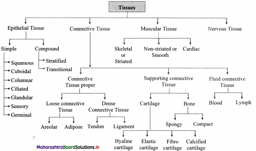
There are four types of animal tissues namely, epithelial, connective, muscular and nervous tissue.
Question 6.
Give the characteristics of epithelial tissue.
Answer:
The characteristics of epithelial tissues are as follows:
- Epithelial tissue forms a covering on inner and outer surface of body and organs.
- The cells of this tissue are compactly arranged with little intercellular matrix.
- The cells rest on a non-cellular basement membrane.
- The epithelial cells are polygonal, cuboidal or columnar in shape.
- A single nucleus is present at the centre or at the base of the cell.
- The tissue is avascular and has a good regeneration capacity.
- The major function of the epithelial tissue is protection. It also helps in absorption, transport, filtration and secretion.
![]()
Question 7.
Name the types of epithelial tissues.
Answer:
The different types of epithelial tissues are as follows:
1. Simple epithelium: Epithelial tissue made up of single layer of cells is known as simple epithelium. Simple epithelium is further classified into:
a. Squamous Epithelium
b. Cuboidal Epithelium
c. Columnar Epithelium
d. Ciliated Epithelium
e. Glandular Epithelium
f. Sensory epithelium
g. Germinal epithelium
2. Compound epithelium: Epithelium composed of several layers is called compound epithelium. Compound epithelium is further classified into:
a. Stratified epithelium
b. Transitional epithelium
Question 8.
What does ‘basement membrane’ signify?
Answer:
Basement membrane is a non – cellular membrane on which the lowermost layer of the epithelium lies.
Question 9.
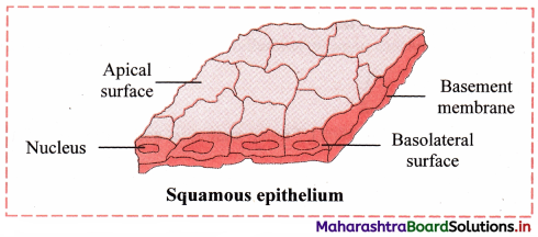
Write a note on squamous epithelium.
Answer:
Squamous epithelium or pavement epithelium:
Location: It is present in blood vessels, alveoli, coelom, etc.
Structure:
- The squamous epithelium is composed of single layer of cells.
- The cells are polygonal in shape, thin and flat, with serrated margin.
- They have centrally placed spherical or oval nucleus.
- They appear like flat tiles when viewed from above, thus, are also called as pavement epithelium.
- Functions: Protection, absorption, transport, filtration and secretion.
Question 10.
Give an account of cuboidal epithelium.
Answer:
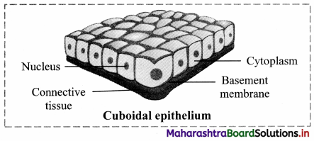
Cuboidal Epithelium:
Location: It is present in the lining of pancreatic ducts, salivary duct, proximal and distal convoluted tubules of nephron, etc.
Structure:
1. The cells are cuboidal in shape.
2. They have a centrally placed, spherical nucleus.
Functions: Absorption and secretion.
![]()
Question 11.
Describe briefly about columnar epithelium.
Answer:
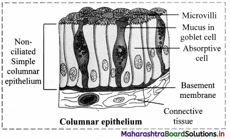
Columnar Epithelium:
Location: It is found in inner lining of intestine, gall bladder, gastric glands, intestinal glands, etc.
Structure:
1. The cells are tall, pillar-like. The inner ends of the cells are narrow while free ends are broad and flat.
2. Nucleus is oval or elliptical in the lower half of the cell.
3. Free surface shows large number of microvilli.
Function: Secretion and absorption.
Question 12.
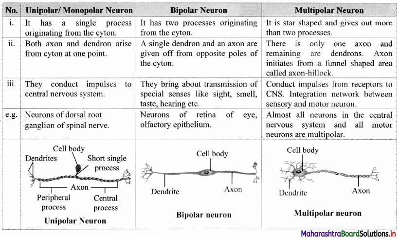
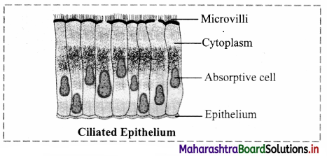
Write a note on ciliated epithelium.
Answer:
Location: It is found in inner lining of buccal cavity of frog, nasal cavity, trachea, oviduct of vertebrates, etc.
Structure:
1. Cells of this tissue are cuboidal or columnar.
2. Free ends of cells are broad while narrow ends rest on a basement membrane.
3. The free ends of the cell show hair-like cilia.
4. The nucleus is oval and placed at basal end of the cell.
Function: To create a movement of materials that comes in contact with the epithelium, in a specific direction. This aids in functions like prevention of entry of foreign particles in the trachea, pushing of the ovum through the oviduct, etc.
Question 13.
What is sensory epithelium? Draw a neat and labelled diagram.
Answer:
Sensory epithelium is composed of a modified form of columnar cells and elongated neurosensory cells. Sensory hairs are present at the free end of these cells.
Function: It perceives external as well as internal stimuli.
Location: It is found in the nose (Olfactory), ear (Auditory hair cells) and eye (photoreceptors).

Question 14.
What is the function of germinal epithelium?
Answer:
The cells of the germinal epithelium divide meiotically to produce haploid gametes, e.g. Lining of seminiferous tubules, inner lining of ovary, etc.
![]()
Question 15.
Explain compound epithelium with a suitable diagram.
Answer:
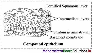
1. Compound epithelium consists of many layers of cells.
2. Only the lowermost layer of this tissue is based on the basement membrane.
3. Types of compound epithelium include:
a. Stratified epithelium: Nucleus is present in stratum germinativum (basal layer).
Cells at free surface become flat and lack nucleus called stratum comeum.
Function: Protection e.g. Epidermis of skin, oesophagus, cornea, vagina, rectum.
b. Transitional epithelium:
Structure of transitional epithelium is same like stratified epithelium.
The cells can undergo a change in their shape and structure depending on degree of stretch. Function: Distension of organ e.g. Urinary bladder
Question 16.
Distinguish between simple epithelium and compound epithelium.
Answer:
| Simple epithelium | Compound epithelium |
| 1. It is made up of single layer of cells. | It is made up of two or more layer of cells. |
| 2. Single layer of cells that rest on the basement membrane. | Only lowermost layer rests on basement membrane |
| 3. It is useful in diffusion, osmosis, filtration, secretion and absorption. | Generally protective in function. It has limited role in absorption. |
| 4. It is generally present in the outer and inner lining of organs, blood vessels etc. | It is present in the epidermis of skin, oesophagus, cornea, vagina, rectum, urinary bladder, etc. |
Question 17.
Identify the type of epithelium found in the following cells/ cell structures:
- Auditory hair cells
- Goblet cells
- Inner lining of gall bladder
- Lining of oviduct of vertebrates
- Urinary bladder
Answer:
- Sensory epithelium
- Glandular epithelium
- Columnar epithelium
- Ciliated epithelium
- Transitional epithelium (Compound epithelium)
![]()
Question 18.
What is connective tissue? Write its characteristics.
Answer:
Connective tissue is the most widely spread tissue in the body which binds, supports and provides strength to other body tissues and organs.
Characteristics:
1. It consists of a variety of cells and fibres which are embedded in the abundant intercellular substance called matrix.
2. It is a highly vascular tissue, except cartilage.
3. The connective tissue is classified on the basis of matrix present, into three types, namely connective tissue proper, supporting connective tissue and fluid connective tissue.
a. Connective tissue proper is further classified as loose connective tissue (e.g. areolar connective tissue and adipose tissue) and dense connective tissue (e.g. ligament and tendon).
b. Supporting connective tissue also called skeletal tissue includes cartilage and bone.
c. Fluid connective tissue includes blood and lymph.
4. Functions: Connective tissue protects the vital organs of the body. It acts as packing material and also helps in healing process.
Question 19.
Name the cells of connective tissue which form fibers.
Answer:
Fibroblasts are the cells of connective tissue which form fibres.
Question 20.
Distinguish between epithelial tissue and connective tissue.
Answer:
| Epithelial tissue | Connective tissue |
| 1. No intercellular space is present between the cells. | Large intercellular space present between the cell. |
| 2. Basement membrane present. | Basement membrane absent. |
| 3. Functions include covering, protection, secretion. | Functions include attachment, support, storage, transportation. |
| 4. It is present in the skin, lung alveoli, kidney tubules, etc. | It is present in tendons, ligament, bone, etc. |
Question 21.
Fill in the blanks.
- Connective tissues are highly vascular, except _______ .
- Supporting connective tissues are also called as ______ .
- Areolar tissue is a type of _______ connective tissue.
Answer:
- cartilage
- skeletal tissue
- loose
Question 22.
With help of neat labelled diagram, describe the structure of areolar connective tissue.
Answer:
Areolar tissue is a loose connective tissue found under the skin, between muscles, bones, around organs, blood vessels and peritoneum. It is composed of fibres and cells.
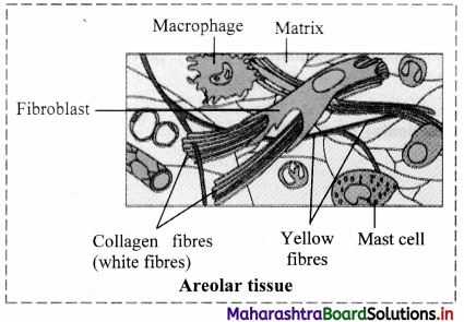
The matrix of areolar tissues contains two types of fibres i.e. white fibres and yellow fibres.
a. White fibres: They are made up of collagen and give tensile strength to the tissue.
b. Yellow fibres: They are made up of elastin and are elastic in nature.
The four different types of cells present in this tissue are as follows:
a. Fibroblast: Large flat cells having branching processes. They produce fibres as well as polysaccharides that form the ground substance or matrix of the tissue.
b. Mast cells: Oval cells that secrete heparin and histamine.
c. Macrophages: Amoeboid, phagocytic cells.
d. Adipocytes (Fat cells): These cells store fat and have eccentric nucleus.
![]()
Question 23.
What is the function of areolar tissue?
Answer:
Areolar tissue acts as packing material, helps in healing process and connects different organs or layers of tissues.
Question 24.
Give the location, structure and function of adipose tissue.
Answer:
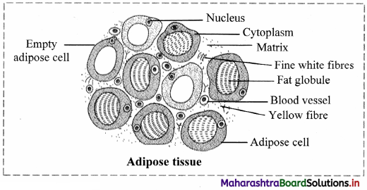
Adipose tissue (adipo = fat):
Location: It is found in association with areolar connective tissue. Adipose tissue is present beneath the skin, around the kidneys and between internal organs.
Structure:
- It contains large number of adipocytes.
- The cells are rounded or polygonal.
- Due to presence of fats stored in the form of droplets in adipocytes, the nucleus is shifted towards the periphery.
- Matrix is less and fibres and blood vessels are few in number.
The adipose tissue is of two types:
a. White adipose tissue:
1. It is opaque due to the presence of large number of adipocytes.
2. It is commonly present in adults.
b. Brown adipose tissue:
It is reddish brown in colour due to the presence of large number of blood vessels.
Functions:
- Adipose tissue is a good insulator, acts as a shock absorber and a good source of energy because it stores fat.
- The tissue is found in the sole and palm region as well as around organs like kidneys.
- The number of fat cells do not decrease on dieting. Once fat cells are formed, they remain constant throughout adult life. Dieting can only reduce the size of the fat cells and not their number.
- A person may generally have 10 – 30 billion fat cells in their body. Obese people can eventually have up to 100 billion fat cells.
Question 25.
State the two types of dense connective tissue.
Answer:
1. Fibres and fibroblasts are compactly arranged in the dense connective tissue.
2. There are two types of dense connective tissue:
a. Dense regular connective tissue: Collagen fibres are arranged in a parallel manner, e.g. Tendons and ligaments.
b. Dense irregular connective tissue: Fibres and fibroblasts are not arranged in an orderly manner, e.g. Dermis of skin.
![]()
Question 26.
Write a short note on tendon.
Answer:
1. Tendons are a type of dense regular connective tissue.
2. Tendons connect skeletal muscles to bones.
3. They contain bundles of white fibres which give tensile strength to the tissue, e.g. Achilles tendon, Hamstring tendon.
Question 27.
Raju is experiencing pain at the back of the ankle and lower calf after a serious injury in a football match. What tissue must he have injured?
Answer:
Pain at the back of the ankle and lower calf indicates an injury to the tendons (dense connective tissue – Achilles tendon).
Question 28.
What are ligaments? Where are ligaments present and what is their function?
Answer:
Ligaments are a type of dense regular connective tissue that are made up of elastic or yellow fibres arranged in regular pattern. These fibres make the ligaments elastic.
Location: Ligaments are present at joints.
Function: Ligaments prevent dislocation of bones.
Question 29.
Identify the labels (X and Y) in the given diagram.
image
Answer:
X: Tendon Y: Ligament
Question 30.
What is supporting connective tissue? What are its types?
Answer:
1. Supporting connective tissue is a type of connective tissue which is characterised by the presence of hard matrix.
2. It is classified into two types i.e., cartilage and bone.
Question 31.
Write a short note on cartilage.
Answer:
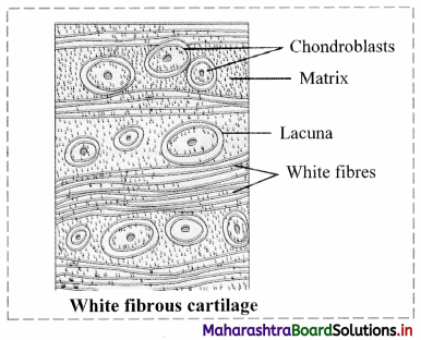
Cartilage is a type of supporting connective tissue. It is a pliable yet tough tissue.
Structure:
- Abundant matrix is delimited by a sheath of collagenous fibres called perichondrium.
- The matrix is called chondrin.
- Below the perichondrium, immature cartilage forming cells called chondroblasts are present.
- Chondroblasts mature and get converted into chondrocytes.
Question 32.
Explain in brief about the various types of cartilages, with the help of a suitable diagram.
Answer:
Cartilage is a type of supporting connective tissue.
Depending upon the nature of the matrix, cartilage is of four types.
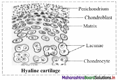
1. Hyaline cartilage: The hyaline cartilage is elastic and compressible in nature.
a. Perichondrium is present in this cartilage.
b. Its matrix is bluish white and gel like.
c. Very fine collage fibres and chondrocytes are present in this cartilage.
Function: It acts as a good shock absorber as well as provides flexibility. It reduces friction. Location: It is found at the end of long bones, epiglottis, trachea, ribs, larynx and hyoid.
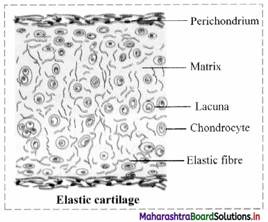
2. Elastic cartilage:
a. The perichondrium is present in elastic cartilage.
b. The matrix contains elastic fibres and chondrocytes are few in numbers.
Function: It gives support and maintains shape of the body part.
Location: It is found in the ear lobe, tip of the nose, etc.
3. Fibrocartilage:
a. The fibrocartilage is the most rigid cartilage.
b. Perichondrium is absent in the fibrocartilage.
c. The matrix contains bundles of collagen fibres and few chondrocytes that are scattered in the fibres.
Function: It maintains position of vertebrae.
Location: Intervertebral discs are made up of fibrocartilage. It is also found at the pubic symphysis.
4. Calcified cartilage:
This type of cartilage becomes rigid due to deposition of salts in the matrix, reducing the flexibility of joints in old age.
e.g. Head of long bones.
![]()
Question 33.
Fill in the blanks by selecting the correct word from the bracket and complete the given paragraph.
(heart, chondrium, peristomium, bone, perichondrium, ossein, pubic symphysis, cartilage, lacunae, chondrocytes)
_______ is a pliable yet tough supporting connective tissue. Its matrix is surrounded by a sheath of
collagenous fibres called _______ . The matrix is called ________ . In chondroblasts mature and get converted into ________ which are enclosed in the ________ in the matrix. This type of connective tissue is generally found in the _______ , ear lobe, etc.
Answer:
Cartilage is a pliable yet tough supporting connective tissue. Its matrix is surrounded by a sheath of collagenous fibres called perichondrium. The matrix is called chondrin. The chondroblasts mature and get converted into chondrocytes which are enclosed in the lacunae in the matrix. This type of connective tissue is generally found in the pubic symphysis, ear lobe, etc.
Question 34.
Distinguish between elastic cartilage and fibrocartilage.
Answer:
| Elastic cartilage | Fibrocartilage |
| 1. Perichondrium is present. | Perichondrium is absent. |
| 2. Very fine collagen fibres and chondrocytes are present in the matrix. | Matrix contains bundles of collagen fibres and few chondrocytes. |
| 3. It is elastic and compressible in nature. | It is the most rigid cartilage. |
| 4. It acts as a good shock absorber and provides flexibility. | It maintains the position of vertebrae. |
Question 35.
Which protein is present in the bone matrix?
Answer:
Ossein is present in the bone matrix.
Question 36.
Based on the presence of matrix classify the bones present in the human body.
Answer:
Based on the presence of matrix there are two types of bones present in the human body:
1. Spongy bones:
Haversian system is absent in these bones.
Rectangular matrix is arranged in the form of trabeculae.
It contains red bone marrow.
2. Compact bones:
Matrix of these bones shows haversian system without any space between the lamellae.
Question 37.
Distinguish between cartilage and bone.
Answer:
| Cartilage | Bone |
| 1. Matrix is covered by a sheath of collagenous fibres called perichondrium. | Matrix is surrounded by an outer tough membrane called periosteum. |
| 2. Cartilage is ilexible. | Bone is rigid. |
| 3. Haversian system is absent. | Haversian system is present in mammalian bones. |
| 4. Matrix is made up of chondrin. | Matrix is made up of ossein. |
![]()
Question 38.
Name the fluid connective tissues present in the body of animals.
Answer:
Blood and lymph are fluid connective tissues present in the body of animals.
Question 39.
Give the characteristics of muscular tissue.
Answer:
- The cells of the muscular tissue are elongated and are called as muscle fibres.
- The muscle fibres are covered by a membrane called sarcolemma.
- The cytoplasm of the muscle cell is called the sarcoplasm.
- Large number of contractile fibrils called myofibrils are present in the sarcoplasm.
- Depending on the type of muscle cells, one or many nuclei may be present.
- Myofibrils are made up of the proteins, actin and myosin.
- Muscle fibres contract and decrease in length on stimulation. Hence, muscular tissue is also known as contractile tissue.
- This tissue is vascular and innervated by nerves.
- Muscle cells contain large number of mitochondria.
Question 40.
Mention the different types of muscles and give their locations.
Answer:
1. Skeletal muscles/Striated muscles/ Voluntary muscles: They are found attached to bones.
2. Smooth / Non-striated muscles/ Involuntary muscles: They are found in the walls of visceral organs and blood vessels.
3. Cardiac muscles: They are found in the wall of the heart or myocardium.
Question 41.
With the help of a neat and labelled diagram, describe the location, structure and function of skeletal muscles.
Answer:
Skeletal muscles are also known as voluntary muscles.
Location: Skeletal muscles are found attached to bones.
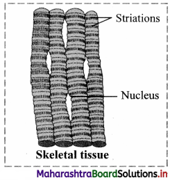
Structure:
1. They consist of large number of fasciculi which are wrapped by a connective tissue sheath called epimysium or fascia. Each individual fasciculus covered by perimysium.
2. Each fasciculus in turn consists of many muscle fibres called myofibers.
3. Each muscle fibre is a syncytial fibre that contains several nuclei.
4. The sarcoplasm (cytoplasm) is surrounded by the sarcolemma (cell membrane).
5. The sarcoplasm contains large number of parallelly arranged myofibrils and hence the nuclei gets shifted to the periphery.
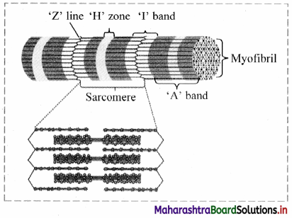
6. Each myofibril is made up of repeated functional units called sarcomeres.
7. Each sarcomere has a dark band called anisotropic of ‘A’ band in the centre. ‘A’ bands are made up of the contractile proteins actin and myosin.
In the centre of the ‘A’ band is the light area called ‘H’ zone or Hensen’s zone.
In the centre of the Hensen’s zone is the ‘M’ line.
On either side of the ‘A’ band are light bands called isotropic or ‘I’ bands. These bands contain only actin.
Adjacent light bands are separated by the ‘Z’ line (Zwischenscheibe line).
The dark and light bands on neighbouring myofibrils correspond with each other to give the muscles a striated appearance.
Functions: Skeletal muscles bring about voluntary movements of the body
Question 42.
Observe the given diagram and answer the questions given below it.
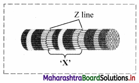
1. What does the given diagram represent?
2. Identify ‘X’ in the given diagram.
Answer:
1. The given diagram represents the myofibril of a muscular tissue.
2. ‘X’ is the sarcomere. It is a repeating unit of contraction within the myofibril.
![]()
Question 43.
Which is the functional unit of skeletal muscles?
Answer:
Sarcomere is the functional unit of skeletal muscles.
Question 44.
What are the different types of skeletal muscles?
Answer:
Skeletal muscles are divided into two types based on the amount of red pigment (myoglobin).
1. Red muscle: It contains very high amount of myoglobin.
2. White muscle: It contains very low amount of myoglobin.
Question 45.
What is myoglobin? What is its function?
Answer:
Myoglobin is an iron containing red coloured pigment found only in muscles. It consists of one haeme and one polypeptide chain. It can carry one molecule of oxygen.
Function: Due to presence of myoglobin, the muscles can obtain their oxygen from two sources, myoglobin and haemoglobin.
Question 46.
Describe the structure, location and function of smooth muscles.
Answer:
Smooth muscles are also known as non-striated, visceral or involuntary muscles.
Structure:
- These muscles are present in the form of sheets or layers.
- Each muscle cell is spindle shaped or fusiform.
- The fibres are unbranched and have a single nucleus that is located centrally.
- The sarcoplasm contains myofibrils which are made up of the contractile proteins – actin and myosin.
- Smooth muscles contain less myosin and more actin.
- Striations are absent, hence smooth muscles are also known as non-striated muscles,
- These muscles are innervated by the autonomous nervous system.
Location:
It is found in walls of visceral organs and blood vessels. Therefore, smooth muscles are also known as visceral muscles.
Function:
Smooth muscles are associated with involuntary movements of the body like peristaltic movement of food through the digestive system.
![]()
Question 47.
Distinguish between smooth muscles and skeletal muscles.
Answer:
| Smooth Muscles | Skeletal Muscles |
| 1. These muscles are found in the walls of visceral organs and blood vessels. | These muscles are found attached to the bone. |
| 2. Each muscle cell is spindle shaped or fusiform and unbranched | They are cylindrical in shape and branched. |
| 3. They have a single, centrally located nucleus. | They contain several nuclei that are shifted to the periphery due to presence of large number of myofibrils. |
| 4. Striations are absent in smooth muscles. | Striations are present in skeletal muscles. |
| 5. They undergo slow and sustained involuntary contractions. | They show quick and strong voluntary contractions. |
| 6. They contain lesser myosin are more actin as compared to skeletal muscles. | They contain more myosin and lesser actin as compared to smooth muscles. |
Question 48.
Describe the structure, location and function of cardiac muscle fibres.
Answer:
Muscles of the cardiac tissue show characteristics of both striated and non-striated muscle fibres.
Structure:
- Sarcolemma is not distinct.
- Uninucleate muscle fibres appear to be multinucleate.
- Adjacent muscle fibres join together to give branched appearance.
- Transverse thickenings of the sarcolemma called intercalated discs form points of adhesion of muscle fibres.
- These junctions allow cardiac muscles to contract as a unit to aid quick transfer of stimulus.
Location:
They are found in the wall of the heart or myocardium.
Function:
Cardiac muscles bring about contraction and relaxation of heart, which helps in circulation of blood throughout the body.
Question 49.
Why is the mammalian heart known as a myogenic heart?
Answer:
The mammalian cardiac muscles are modified and are capable of generating an impulse on their own. Hence, the mammalian heart known as a myogenic heart.
Question 50.
What is a neurogenic heart?
Answer:
In some animals, the cardiac muscles need neural stimulus in order to initiate a contraction. Such a heart is known as a neurogenic heart.
Question 51.
Describe the characteristics of the nervous tissue.
Answer:
The characteristics of the nervous tissue are as follows:
- The nervous tissue is made up of nerve cells (neurons) and neuroglia.
- Intracellular matrix is absent in the neural tissue.
- The neurons are the structural and functional units of the nervous system.
a. They are impulse generating and impulse conducting units which bring about quick communication within the body.
b. Excitability is the change in action potential of the neuronal membrane on receiving external stimulus.
c. Conductivity helps the neurons to carry a wave of impulse from the dendron to the axon (processes of neuron).
Question 52.
What are neuroglial cells?
Answer:
Neuroglial cells are non-nervous supporting cells that fill in the inter-neuronal space and are capable of regeneration and division.
![]()
Question 53.
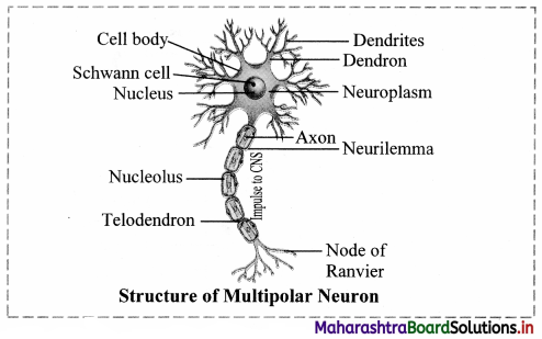
Describe the structure of a neuron.
Answer:
A neuron is the structural and functional unit of the nervous tissue. A neuron is made up of cyton or cell body and cytoplasmic extensions or processes.
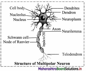
1. Cyton:
The cyton or cell body contains granular cytoplasm called neuroplasm and a centrally placed nucleus. The neuroplasm contains mitochondria, Golgi apparatus, RER and Nissl’s granules.
2. Cytoplasmic extensions or processes:
(a) Dendron: They are short, unbranched processes.
The fine branches of a dendron are called dendrites.
Dendrites carry an impulse towards the cyton.
(b) Axon: It is a single, elongated and cylindrical process.
- The axon is bound by the axolemma.
- The protoplasm or axoplasm contains large number of mitochondria and neurofibrils.
- The axon is enclosed in a fatty sheath called the myelin sheath and the outer covering of the myelin sheath is the neurilemma.
- Both the myelin sheath and the neurilemma are parts of the Schwann cell. The myelin sheath is absent at intervals along the axon at the Node of Ranvier.
- The fine branching structure at the end of the axon (terminal arborization) is called telodendron.
Question 54.
Is a neuron capable of regeneration? Why?
Answer:
No, a neuron is not capable of regeneration because it lacks a centriole.
Question 55.
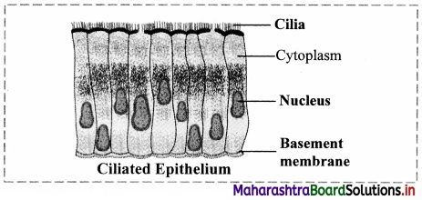
How are neurons classified on the basis of their functions?
Answer:
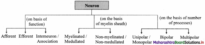
Neurons are classified into three types based on their functions:
1. Afferent neuron (Sensory neuron):
Function: It carries impulses from sense organ to the central nervous system (CNS).
Location: It is found in the dorsal root of the spinal cord.
2. Efferent Neuron (Motor neuron):
Function: It carries impulses from CNS to effector organs.
Location: It is found in the ventral root of the spinal cord.
3. Interneuron or association neuron:
Function: They perfonn processing, integration of sensory impulses and activate appropriate motor neuron to generate motor impulse.
Location: These are located between sensory and motor neurons.
![]()
Question 56.
How are neurons classified depending on the presence or absence of myelin sheath?
Answer:
Depending on the presence or absence of myelin sheath neurons are classified into two types:
1. Myelinated nerve fibre/ medullated nerve fibres: These nerve fibres have an insulating fatty layer called myelin sheath around the axon. This makes the fibre appear white in colour.
2. Non-myelinated/ non-medullated nerve fibres: These nerve fibres lack myelin sheath. The fibres are grey in colour due to absence of myelin sheath.
Question 57.
Conduction of nerve impulse occurs at a faster rate in medullated nerve fibre.
Answer:
- Medullated nerve fibres have myelin sheath around the axon. This myelin sheath is secreted by Schwann cells.
- Myelin sheath prevents the loss of the impulse during conduction.
- Myelin sheath is not continuous. It is interrupted at regular intervals by nodes of Ranvier.
- The nerve impulse jumps from one node to the next and travels faster at these nodes. Such transmission of impulse is called salutatory conduction.
- Therefore, conduction of nerve impulse occurs at a faster rate in medullated nerve fibre.
Question 58.
Compare and contrast between the different types of neurons based on the number of processes given out from the cyton. Draw diagrams.
Answer:
Question 59.
Match the following:
| ‘A’ Group | ‘B’ Group |
| 1. Muscle | (a) Perichondrium |
| 2. Bone | (b) Sarcolemma |
| 3. Nerve cell | (c) Periosteum |
| 4. Cartilage | (d) Neurilemma |
Answer:
| ‘A’ Group | B’ Group |
| 1. Muscle | (b) Sarcolemma |
| 2. Bone | (c) Periosteum |
| 3. Nerve cell | (d) Neurilemma |
| 4. Cartilage | (a) Perichondrium |
![]()
Question 60.
Explain the functions of the different types of epithelial cells.
Answer:
- Epithelial tissue – Protection, secretion, absorption, excretion and filtration.
- Connective tissue – Provides strength to body tissues and organs, protects vital organs, acts as packing material, helps in healing
- Muscular tissue – Movement of body parts and locomotion.
- Nervous tissue – Control and coordination by nerve impulse.
61. Correct the given figures given and write a note.
Question 1.
Answer:
For description of ciliated epithelium: It is found in inner lining of buccal cavity of frog, nasal cavity, trachea, oviduct of vertebrates, etc.
Structure:
1. Cells of this tissue are cuboidal or columnar.
2. Free ends of cells are broad while narrow ends rest on a basement membrane.
3. The free ends of the cell show hair-like cilia.
4. The nucleus is oval and placed at basal end of the cell.
Function: To create a movement of materials that comes in contact with the epithelium, in a specific direction. This aids in functions like prevention of entry of foreign particles in the trachea, pushing of the ovum through the oviduct, etc.
Question 2.
Answer:
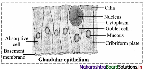
Glandular Epithelium:
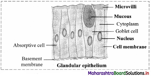
For description of Glandular Epithelium: Structure:
1. The cells of the glandular epithelium can be columnar, cuboidal or pyramidal in shape.
2. The nucleus of these cells is large and situated towards the base.
3. Secretory granules are present in the cell cytoplasm.
4. Glands consist of glandular epithelium. The glands may be either unicellular (goblet cells of intestine) or multicellular (salivary gland), depending on the number of cells.
5. Types: Depending on the mode of secretion, multicellular glands can be further classified as duct bearing glands (exocrine glands) ad ductless glands (endocrine glands).
a. Exocrine glands: These glands pour their secretions at a specific site. e.g. salivary gland, sweat gland, etc.
b. Endocrine glands: These glands release their secretions directly into the blood stream, e.g. thyroid gland, pituitary gland, etc.
vi. Function: Glandular epithelium secretes mucus to trap the dust particles, lubricate the inner surface of respiratory and digestive tracts, secrete enzymes and hormones, etc.
Heterocrine glands
1. Heterocrine glands or composite glands have both exocrine and endocrine function.
2. Pancreas is called a heterocrine gland because it secretes the hormone insulin into
![]()
Question 3.
Answer:
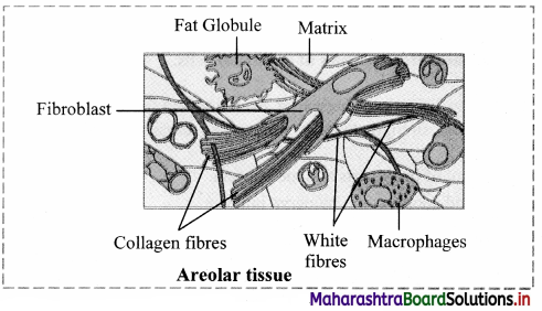
For description and correct diagram of Areolar tissue: Areolar tissue is a loose connective tissue found under the skin, between muscles, bones, around organs, blood vessels and peritoneum. It is composed of fibres and cells.
The matrix of areolar tissues contains two types of fibres i.e. white fibres and yellow fibres.
a. White fibres: They are made up of collagen and give tensile strength to the tissue.
b. Yellow fibres: They are made up of elastin and are elastic in nature.
The four different types of cells present in this tissue are as follows:
a. Fibroblast: Large flat cells having branching processes. They produce fibres as well as polysaccharides that form the ground substance or matrix of the tissue.
b. Mast cells: Oval cells that secrete heparin and histamine.
c. Macrophages: Amoeboid, phagocytic cells.
d. Adipocytes (Fat cells): These cells store fat and have eccentric nucleus.
Question 4.
Answer:
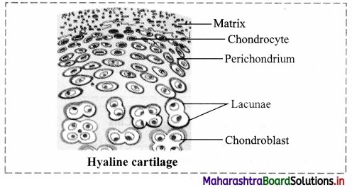
For description and correct diagram of Hyaline cartilage: The hyaline cartilage is elastic and compressible in nature.
a. Perichondrium is present in this cartilage.
b. Its matrix is bluish white and gel like.
c. Very fine collage fibres and chondrocytes are present in this cartilage.
Function: It acts as a good shock absorber as well as provides flexibility. It reduces friction. Location: It is found at the end of long bones, epiglottis, trachea, ribs, larynx and hyoid.
Question 5.
Answer:
For description and correct diagram of Multipolar Neuron: A neuron is the structural and functional unit of the nervous tissue. A neuron is made up of cyton or cell body and cytoplasmic extensions or processes.
1. Cyton:
The cyton or cell body contains granular cytoplasm called neuroplasm and a centrally placed nucleus. The neuroplasm contains mitochondria, Golgi apparatus, RER and Nissl’s granules.
2. Cytoplasmic extensions or processes:
The fine branches of a dendron are called dendrites. Dendrites carry an impulse towards the cyton.
a. Dendron: They are short, unbranched processes.
b. Axon: It is a single, elongated and cylindrical process.
- The axon is bound by the axolemma.
- The protoplasm or axoplasm contains large number of mitochondria and neurofibrils.
- The axon is enclosed in a fatty sheath called the myelin sheath and the outer covering of the myelin sheath is the neurilemma. Both the myelin sheath and the neurilemma are parts of the Schwann cell. The myelin sheath is absent at intervals along the axon at the Node of Ranvier.
- The fine branching structure at the end of the axon (terminal arborization) is called telodendron.
- Nissl’s granules are large granular bodies, found in neurons. These granules are made up of rough endoplasmic reticulum (RER) and free ribosomes (site of protein synthesis).
- It was named after Franz Nissl, a German neuropathologist who invented the Nissl staining method.
![]()
Question 6.
Is a neuron capable of regeneration? Why?
Answer:
No, a neuron is not capable of regeneration because it lacks a centriole.
Question 7.
How are neurons classified on the basis of their functions?
Answer:
Neurons are classified into three types based on their functions:
1. Afferent neuron (Sensory neuron):
Function: It carries impulses from sense organ to the central nervous system (CNS).
Location: It is found in the dorsal root of the spinal cord.
2. Efferent Neuron (Motor neuron):
Function: It carries impulses from CNS to effector organs.
Location: It is found in the ventral root of the spinal cord.
3. Interneuron or association neuron:
Function: They perfonn processing, integration of sensory impulses and activate appropriate motor neuron to generate motor impulse.
Location: These are located between sensory and motor neurons.
Question 62.
Apply Your Knowledge:
Question 1.
Students were asked to observe various tissues under a microscope during their college practical. The teacher explained the various types of tissues. While observing the tissues students had some doubts. They approached the teacher regarding their doubts in practical.
1. How are the skeletal muscle fibres and smooth muscle fibres identified based on their structure?
2. Which type of muscles are found in the myocardium?
Answer:
1. Skeletal muscles are striated muscles as it shows cross-striations in the form of light and dark bands whereas smooth muscles are without striations, thus can be differentiated.
2. Cardiac muscle fibres are found in the myocardium.
![]()
Question 63.
Quick Review
Question 64.
Exercise:
Question 1.
Define the following terms:
1. Germ cells
2. Somatic cells
Answer:
1. Germ cells:
The sperm and the ova are known as germ cells. They are related to reproductive system.
2. Somatic cells:
All body cells except the sperm and the ova are called as somatic cells.
![]()
Question 2.
What is an organ system? Give example.
Answer:
Number of organs combine together to form an organ-system, e.g. Respiratory system.
Question 3.
Enlist the different types of animal tissues.
Answer:
There are four types of animal tissues namely, epithelial, connective, muscular and nervous tissue.
1. Marie Francois Xavier Bichat (1771- 1802), French anatomist and pathologist discovered tissue. He was known as ‘Father of Histology’.
2. M. Bichat worked without a microscope, yet he distinguished 21 types of elementary tissues from which the organs of the human body are composed.
Question 4.
Define histology.
Answer:
The study of structure and arrangement of tissue is called histology.
Question 5.
Give one example each of exocrine and endocrine gland.
Answer:
Types: Depending on the mode of secretion, multicellular glands can be further classified as duct bearing glands (exocrine glands) ad ductless glands (endocrine glands).
a. Exocrine glands: These glands pour their secretions at a specific site. e.g. salivary gland, sweat gland, etc.
b. Endocrine glands: These glands release their secretions directly into the blood stream, e.g. thyroid gland, pituitary gland, etc.
Question 6.
Define.
1. Exocrine glands
2. Endocrine glands
Answer:
1. Exocrine glands: These glands pour their secretions at a specific site. e.g. salivary gland, sweat gland, etc.
2. Endocrine glands: These glands release their secretions directly into the blood stream, e.g. thyroid gland, pituitary gland, etc.
Question 7.
Describe with neat and labelled diagram squamous epithelium.
Answer:
Squamous epithelium or pavement epithelium:
Location: It is present in blood vessels, alveoli, coelom, etc.
Structure:
- The squamous epithelium is composed of single layer of cells.
- The cells are polygonal in shape, thin and flat, with serrated margin.
- They have centrally placed spherical or oval nucleus.
- They appear like flat tiles when viewed from above, thus, are also called as pavement epithelium. Functions: Protection, absorption, transport, filtration and secretion.
![]()
Question 8.
Name the type of muscle fibres forming the inner lining of the intestine and gastric glands.
Answer:
Columnar Epithelium:
Location: It is found in inner lining of intestine, gall bladder, gastric glands, intestinal glands, etc.
Structure:
1. The cells are tall, pillar-like. The inner ends of the cells are narrow while free ends are broad and flat.
2. Nucleus is oval or elliptical in the lower half of the cell.
3. Free surface shows large number of microvilli.
Function: Secretion and absorption.
Question 9.
Write the functions of different types of cell junctions.
Answer:
1. Cell junctions: The epithelial cells are connected to each other laterally as well as to the basement
membrane by junctional complexes called cell junctions.
2. The different types of cell junctions are as follows:
a. Gap Junctions (GJs): These are intercellular connections that allow the passage of ions and small molecules between cells as well as exchange of chemical messages between cells.
b. Adherens Junctions (AJs): They are involved in various signalling pathways and transcriptional regulations.
c. Desmosomes (Ds): They provide mechanical strength to epithelial tissue, cardiac muscles and meninges.
d. Hemidesmosomes (HDs): They allow the cells to strongly adhere to the underlying basement membrane. These junctions help maintain tissue homeostasis by signalling.
e. Tight junctions (TJs): These junctions maintain cell polarity, prevent lateral diffusion of proteins and ions.
Question 10.
Where is ciliated epithelium located?
Answer:
Location: It is found in inner lining of buccal cavity of frog, nasal cavity, trachea, oviduct of vertebrates, etc.
Structure:
1. Cells of this tissue are cuboidal or columnar.
2. Free ends of cells are broad while narrow ends rest on a basement membrane.
3. The free ends of the cell show hair-like cilia.
4. The nucleus is oval and placed at basal end of the cell.
Function: To create a movement of materials that comes in contact with the epithelium, in a specific direction. This aids in functions like prevention of entry of foreign particles in the trachea, pushing of the ovum through the oviduct, etc.
Question 11.
Why squamous epithelium is also called pavement epithelium?
Answer:
They appear like flat tiles when viewed from above, thus, are also called as pavement epithelium. Functions: Protection, absorption, transport, filtration and secretion.
![]()
Question 12.
Dhruvi met with an accident and has temporarily lost her ability to perceive external auditory stimuli. Which tissue must be affected?
Answer:
Sensory epithelium is composed of a modified form of columnar cells and elongated neurosensory cells. Sensory hairs are present at the free end of these cells.
Function: It perceives external as well as internal stimuli.
Location: It is found in the nose (Olfactory), ear (Auditory hair cells) and eye (photoreceptors).
Question 13.
Write names of any four types of simple epithelium.
Answer:
Simple epithelium: Epithelial tissue made up of single layer of cells is known as simple epithelium. Simple epithelium is further classified into:
a. Squamous Epithelium
b. Cuboidal Epithelium
c. Columnar Epithelium
d. Ciliated Epithelium
Question 14.
Write a short note on types of glandular epithelium.
Answer:
Types: Depending on the mode of secretion, multicellular glands can be further classified as duct bearing glands (exocrine glands) ad ductless glands (endocrine glands).
a. Exocrine glands: These glands pour their secretions at a specific site. e.g. salivary gland, sweat gland, etc.
b. Endocrine glands: These glands release their secretions directly into the blood stream, e.g. thyroid gland, pituitary gland, etc.
Question 15.
Describe the structure, function and location of columnar epithelium.
Answer:
Columnar Epithelium:
Location: It is found in inner lining of intestine, gall bladder, gastric glands, intestinal glands, etc.
Structure:
1. The cells are tall, pillar-like. The inner ends of the cells are narrow while free ends are broad and flat.
2. Nucleus is oval or elliptical in the lower half of the cell.
3. Free surface shows large number of microvilli.
Function: Secretion and absorption.
Question 16.
Give the location and function of:
1. Cuboidal epithelium
2. Glandular epithelium
Answer:
1. Cuboidal Epithelium:
Location: It is present in the lining of pancreatic ducts, salivary duct, proximal and distal convoluted tubules of nephron, etc.
Structure:
1. The cells are cuboidal in shape.
2. They have a centrally placed, spherical nucleus.
Functions: Absorption and secretion.
2. Structure:
1. The cells of the glandular epithelium can be columnar, cuboidal or pyramidal in shape.
2. The nucleus of these cells is large and situated towards the base.
3. Secretory granules are present in the cell cytoplasm.
4. Glands consist of glandular epithelium. The glands may be either unicellular (goblet cells of intestine) or multicellular (salivary gland), depending on the number of cells.
5. Types: Depending on the mode of secretion, multicellular glands can be further classified as duct bearing glands (exocrine glands) ad ductless glands (endocrine glands).
a. Exocrine glands: These glands pour their secretions at a specific site. e.g. salivary gland, sweat gland, etc.
b. Endocrine glands: These glands release their secretions directly into the blood stream, e.g. thyroid gland, pituitary gland, etc.
6. Function: Glandular epithelium secretes mucus to trap the dust particles, lubricate the inner surface of respiratory and digestive tracts, secrete enzymes and hormones, etc.
Heterocrine glands:
1. Heterocrine glands or composite glands have both exocrine and endocrine function.
2. Pancreas is called a heterocrine gland because it secretes the hormone insulin into blood which is an endocrine function and enzymes into digestive tract which is an exocrine function.
![]()
Question 17.
With the help of suitable diagram explain compound epithelium.
Answer:
1. Compound epithelium consists of many layers of cells.
2. Only the lowermost layer of this tissue is based on the basement membrane.
3. Types of compound epithelium include:
a. Stratified epithelium: Nucleus is present in stratum germinativum (basal layer).
Cells at free surface become flat and lack nucleus called stratum comeum.
Function: Protection e.g. Epidermis of skin, oesophagus, cornea, vagina, rectum.
b. Transitional epithelium:
Structure of transitional epithelium is same like stratified epithelium.
The cells can undergo a change in their shape and structure depending on degree of stretch. Function: Distension of organ e.g. Urinary bladder
Question 18.
Ciliated epithelium is found in the upper respiratory tract.
Answer:
Function: To create a movement of materials that comes in contact with the epithelium, in a specific direction. This aids in functions like prevention of entry of foreign particles in the trachea, pushing of the ovum through the oviduct, etc.
Question 19.
Give any four characteristics of connective tissue.
Answer:
Characteristics:
1. It consists of a variety of cells and fibres which are embedded in the abundant intercellular substance called matrix.
2. It is a highly vascular tissue, except cartilage.
3. The connective tissue is classified on the basis of matrix present, into three types, namely connective tissue proper, supporting connective tissue and fluid connective tissue.
a. Connective tissue proper is further classified as loose connective tissue (e.g. areolar connective tissue and adipose tissue) and dense connective tissue (e.g. ligament and tendon).
b. Supporting connective tissue also called skeletal tissue includes cartilage and bone.
c. Fluid connective tissue includes blood and lymph.
![]()
Question 20.
What is tendon?
Answer:
Tendons are a type of dense regular connective tissue.
Question 21.
What is a ligament?
Answer:
Ligaments are a type of dense regular connective tissue that are made up of elastic or yellow fibres arranged in regular pattern. These fibres make the ligaments elastic.
Location: Ligaments are present at joints.
Function: Ligaments prevent dislocation of bones.
Question 22.
Write a note on hyaline cartilage.
Answer:
Hyaline cartilage: The hyaline cartilage is elastic and compressible in nature.
a. Perichondrium is present in this cartilage.
b. Its matrix is bluish white and gel like.
c. Very fine collage fibres and chondrocytes are present in this cartilage.
Function: It acts as a good shock absorber as well as provides flexibility. It reduces friction.
Location: It is found at the end of long bones, epiglottis, trachea, ribs, larynx and hyoid.
Question 23.
Give two examples of tendons.
Answer:
1. Achilles tendons (Calcaneal tendons) connect the calf muscles to the heel bone.
2. When the calf muscles flex, the Achilles tendon pulls on the heel. This movement allows us to stand on
our toes.
3. Generally, a pain at the back of ankle or lower calf may signal a problem with an Achilles Tendon.
Athletes who participate in track and field may face Achilles tendon injury, i iv. The Achilles tendon is the largest and strongest tendon in the body.
Question 24.
Write a short note on mammalian bone.
Answer:
Explain histological structure of mammalian bone.
a. The bone is characterised by hard matrix called ossein which is made up of mineral salt hydroxy apatite (Ca10 (P04)6 (OH)2).
b. An outer tough membrane called periosteum encloses the matrix.
c. Blood vessels and nerves pierce through the periosteum.
d. The matrix is arranged in the form of concentric layers called lamellae.
e. Each lamella contains fluid filled cavities called lacunae from which fine canals called canaliculi radiate.
f. The canaliculi of adjacent lamellae connect with each other as they traverse through the matrix.
g. Active bone cells called osteoblasts and inactive bone cells called osteocytes are present in the
lacunae.
h. The mammalian bone shows the peculiar haversian system.
i. The haversian canal encloses an artery, vein and
![]()
Question 25.
Describe briefly about various types of cartilages, with the help of suitable diagram.
Answer:
Cartilage is a type of supporting connective tissue.
Depending upon the nature of the matrix, cartilage is of four types.
1. Hyaline cartilage: The hyaline cartilage is elastic and compressible in nature.
a. Perichondrium is present in this cartilage.
b. Its matrix is bluish white and gel like.
c. Very fine collage fibres and chondrocytes are present in this cartilage.
Function: It acts as a good shock absorber as well as provides flexibility. It reduces friction. Location: It is found at the end of long bones, epiglottis, trachea, ribs, larynx and hyoid.
2. Elastic cartilage:
a. The perichondrium is present in elastic cartilage.
b. The matrix contains elastic fibres and chondrocytes are few in numbers.
Function: It gives support and maintains shape of the body part.
Location: It is found in the ear lobe, tip of the nose, etc.
3. Fibrocartilage:
a. The fibrocartilage is the most rigid cartilage.
b. Perichondrium is absent in the fibrocartilage.
c. The matrix contains bundles of collagen fibres and few chondrocytes that are scattered in the fibres.
Function: It maintains position of vertebrae.
Location: Intervertebral discs are made up of fibrocartilage. It is also found at the pubic symphysis.
Question 26.
Sharada saw that her grandmother is suffering from joint pain and reduced joint flexibility. What tissue is associated with this problem and why does it occur?
Answer:
Calcified cartilage:
This type of cartilage becomes rigid due to deposition of salts in the matrix, reducing the flexibility of joints in old age.
e.g. Head of long bones.
Question 27.
Differentiate between the following:
1. Bone and Cartilage
2. Epithelial tissue and Connective tissue
3. Hyaline cartilage and Fibrocartilage
Answer:
1. Hyaline cartilage: The hyaline cartilage is elastic and compressible in nature.
a. Perichondrium is present in this cartilage.
b. Its matrix is bluish white and gel like.
c. Very fine collage fibres and chondrocytes are present in this cartilage.
Function: It acts as a good shock absorber as well as provides flexibility. It reduces friction.
Location: It is found at the end of long bones, epiglottis, trachea, ribs, larynx and hyoid.
2. Fibrocartilage:
a. The fibrocartilage is the most rigid cartilage.
b. Perichondrium is absent in the fibrocartilage.
c. The matrix contains bundles of collagen fibres and few chondrocytes that are scattered in the fibres.
Function: It maintains position of vertebrae.
Location: Intervertebral discs are made up of fibrocartilage. It is also found at the pubic symphysis.
![]()
Question 28.
Write a note on the structure and location of cartilage.
Answer:
Cartilage is a type of supporting connective tissue. It is a pliable yet tough tissue.
Structure:
1. Abundant matrix is delimited by a sheath of collagenous fibres called perichondrium.
2. The matrix is called chondrin.
3. Below the perichondrium, immature cartilage forming cells called chondroblasts are present.
4. Chondroblasts mature and get converted into chondrocytes.Chondrocytes are scattered in the matrix and are enclosed in the lacunae Each lacuna contains 2 to 8 chondrocytes.
5. It forms the endoskeleton of cartilaginous fishes like shark.
6. It is widely distributed in vertebrate animals
Question 29.
Mention the types of:
1. Fluid connective tissue
2. Supporting connective tissue
Answer:
1. Blood and lymph are fluid connective tissues present in the body of animals.
2. It is classified into two types i.e., cartilage and bone.
Question 30.
Name the protein found in bone matrix.
Answer:
Ossein is present in the bone matrix.
Question 31.
With a neat and labelled diagram explain the structure of adipose tissue.
Answer:
Structure:
- It contains large number of adipocytes.
- The cells are rounded or polygonal.
- Due to presence of fats stored in the form of droplets in adipocytes, the nucleus is shifted towards the periphery.
- Matrix is less and fibres and blood vessels are few in number.
- The adipose tissue is of two types:
Question 32.
Sketch and label multipolar neuron.
Answer:
A neuron is the structural and functional unit of the nervous tissue. A neuron is made up of cyton or cell body and cytoplasmic extensions or processes.
Cyton:
The cyton or cell body contains granular cytoplasm called neuroplasm and a centrally placed nucleus. The neuroplasm contains mitochondria, Golgi apparatus, RER and Nissl’s granules.
![]()
Question 33.
Enlist the characteristics of muscular tissue.
Answer:
- The cells of the muscular tissue are elongated and are called as muscle fibres.
- The muscle fibres are covered by a membrane called sarcolemma.
- The cytoplasm of the muscle cell is called the sarcoplasm.
- Large number of contractile fibrils called myofibrils are present in the sarcoplasm.
- Depending on the type of muscle cells, one or many nuclei may be present.
- Myofibrils are made up of the proteins, actin and myosin.
- Muscle fibres contract and decrease in length on stimulation. Hence, muscular tissue is also known as contractile tissue.
- This tissue is vascular and innervated by nerves.
- Muscle cells contain large number of mitochondria.
Question 34.
Describe in detail, the structure of skeletal muscle fibre.
Answer:
Structure:
- They consist of large number of fasciculi which are wrapped by a connective tissue sheath called epimysium or fascia. Each individual fasciculus covered by perimysium.
- Each fasciculus in turn consists of many muscle fibres called myofibers.
- Each muscle fibre is a syncytial fibre that contains several nuclei.
- The sarcoplasm (cytoplasm) is surrounded by the sarcolemma (cell membrane).
- The sarcoplasm contains large number of parallelly arranged myofibrils and hence the nuclei gets shifted to the periphery.
- Each myofibril is made up of repeated functional units called sarcomeres.
- Each sarcomere has a dark band called anisotropic of ‘A’ band in the centre. ‘A’ bands are made up of the contractile proteins actin and myosin.
- In the centre of the ‘A’ band is the light area called ‘H’ zone or Hensen’s zone.
- In the centre of the Hensen’s zone is the ‘M’ line.
- On either side of the ‘A’ band are light bands called isotropic or ‘I’ bands. These bands contain only actin.
Adjacent light bands are separated by the ‘Z’ line (Zwischenscheibe line). - The dark and light bands on neighbouring myofibrils correspond with each other to give the muscles a striated appearance.
Functions: Skeletal muscles bring about voluntary movements of the body
Question 35.
Describe in detail the location, structure and functions of smooth muscles.
Answer:
Smooth muscles are also known as non-striated, visceral or involuntary muscles.
Structure:
- These muscles are present in the form of sheets or layers.
- Each muscle cell is spindle shaped or fusiform.
- The fibres are unbranched and have a single nucleus that is located centrally.
- The sarcoplasm contains myofibrils which are made up of the contractile proteins – actin and myosin.
- Smooth muscles contain less myosin and more actin.
- Striations are absent, hence smooth muscles are also known as non-striated muscles, vii These muscles are innervated by the autonomous nervous system.
Location:
It is found in walls of visceral organs and blood vessels. Therefore, smooth muscles are also known as visceral muscles.
Function:
Smooth muscles are associated with involuntary movements of the body like peristaltic movement of food through the digestive system.
![]()
Question 36.
What is the importance of myoglobin?
Answer:
Myoglobin is an iron containing red coloured pigment found only in muscles. It consists of one haeme and one polypeptide chain. It can carry one molecule of oxygen.
Function: Due to presence of myoglobin, the muscles can obtain their oxygen from two sources, myoglobin and haemoglobin.
Question 37.
Give the characteristics of nervous tissue.
Answer:
The characteristics of the nervous tissue are as follows:
1. The nervous tissue is made up of nerve cells (neurons) and neuroglia.
2. Intracellular matrix is absent in the neural tissue.
3. The neurons are the structural and functional units of the nervous system.
a. They are impulse generating and impulse conducting units which bring about quick communication within the body.
b. Excitability is the change in action potential of the neuronal membrane on receiving external stimulus.
c. Conductivity helps the neurons to carry a wave of impulse from the dendron to the axon (processes of neuron).
Question 38.
Describe location, structure and functions of cardiac muscles.
Answer:
Muscles of the cardiac tissue show characteristics of both striated and non-striated muscle fibres.
Structure:
1. Sarcolemma is not distinct.
2. Uninucleate muscle fibres appear to be multinucleate.
3. Adjacent muscle fibres join together to give branched appearance.
4. Transverse thickenings of the sarcolemma called intercalated discs form points of adhesion of muscle fibres. These junctions allow cardiac muscles to contract as a unit to aid quick transfer of stimulus.
Location:
They are found in the wall of the heart or myocardium.
Function:
Cardiac muscles bring about contraction and relaxation of heart, which helps in circulation of blood throughout the body.
Question 39.
Explain in detail the structure of neuron.
Answer:
A neuron is the structural and functional unit of the nervous tissue. A neuron is made up of cyton or cell body and cytoplasmic extensions or processes.
1. Cyton:
The cyton or cell body contains granular cytoplasm called neuroplasm and a centrally placed nucleus. The neuroplasm contains mitochondria, Golgi apparatus, RER and Nissl’s granules.
2. Cytoplasmic extensions or processes:
The fine branches of a dendron are called dendrites. Dendrites carry an impulse towards the cyton.
a. Dendron: They are short, unbranched processes.
b. Axon: It is a single, elongated and cylindrical process.
The axon is bound by the axolemma.
The protoplasm or axoplasm contains large number of mitochondria and neurofibrils.
The axon is enclosed in a fatty sheath called the myelin sheath and the outer covering of the myelin sheath is the neurilemma. Both the myelin sheath and the neurilemma are parts of the Schwann cell. The myelin sheath is absent at intervals along the axon at the Node of Ranvier.
The fine branching structure at the end of the axon (terminal arborization) is called telodendron.
1. Nissl’s Granules
2. Nissl’s granules are large granular bodies, found in neurons. These granules are made up of rough endoplasmic reticulum (RER) and free ribosomes (site of protein synthesis).
3. It was named after Franz Nissl, a German neuropathologist who invented the Nissl staining method.
![]()
Question 40.
What is a sarcomere?
Answer:
Sarcomere is the functional unit of skeletal muscles.
Question 41.
What is the difference between myogenic and neurogenic heart?
Answer:
The mammalian cardiac muscles are modified and are capable of generating an impulse on their own. Hence, the mammalian heart known as a myogenic heart.
In some animals, the cardiac muscles need neural stimulus in order to initiate a contraction. Such a heart is known as a neurogenic heart.
Question 42.
Differentiate between neurons on the basis of their functions.
Answer:
Based on the presence of matrix there are two types of bones present in the human body:
1. Spongy bones:
Haversian system is absent in these bones.
Rectangular matrix is arranged in the form of trabeculae.
It contains red bone marrow.
2. Compact bones:
Matrix of these bones shows haversian system without any space between the lamellae.
Question 65.
Multiple Choice Questions
Question 1.
Collagen fibres in the connective tissue are
(A) white
(B) yellow
(C) red
(D) colourless
Answer:
(A) white
![]()
Question 2.
The yellow fibres are chemically composed of
(A) myosin
(B) elastin
(C) collagen
(D) actin
Answer:
(B) elastin
Question 3.
The tissue that stores fats in mammals is
(A) adipose tissue
(B) areolar tissue
(C) nervous tissue
(D) muscular tissue
Answer:
(A) adipose tissue
Question 4.
Ligaments join
(A) muscles to bones
(B) nerves to muscles
(C) skin to muscles
(D) bones to bones
Answer:
(D) bones to bones
Question 5.
The sheath of collagenous fibres, covering the cartilage is known as
(A) perichondrium
(B) periosteum
(C) endosteum
(D) peritoneum
Answer:
(A) perichondrium
Question 6.
A cartilage is formed by
(A) osteoblast
(B) fibroblast
(C) chondrocytes
(D) osteocytes
Answer:
(C) chondrocytes
![]()
Question 7.
The most rigid cartilage is the
(A) fibrous cartilage
(B) elastic cartilage
(C) hyaline cartilage
(D) simple cartilage
Answer:
(A) fibrous cartilage
Question 8.
Active bone cells are called
(A) osteoblast
(B) osteocytes
(C) osteoclasts
(D) osteoporosis
Answer:
(A) osteoblast
Question 9.
Canaliculi is the
(A) space between lamellae
(B) outer tough membrane of the bone
(C) fibres joining adjacent neurons
(D) fine canals that radiate from each lacuna
Answer:
(D) fine canals that radiate from each lacuna
Question 10.
Which of the following is the contractile protein of a muscle?
(A) Tubulin
(B) Myosin
(C) Tropomyosin
(D) Trypsin
Answer:
(B) Myosin
Question 11.
Cytoplasm of muscle cell is called
(A) sarcolemma
(B) neuroplasm
(C) axoplasm
(D) sarcoplasm
Answer:
(D) sarcoplasm
![]()
Question 12.
The structural and functional unit of muscle fibres is
(A) sarcomere
(B) sarcolemma
(C) sarcoplasm
(D) myofibril
Answer:
(A) sarcomere
Question 13.
Dark bands present in the sarcomere are called
(A) ‘A’ band
(B) ‘Z’ lines
(C) ‘H’ line
(D) ‘I’ band
Answer:
(A) ‘A’ band
Question 14.
Nissl’s granules are found in
(A) cartilage cells
(B) nerve cells
(C) muscle cells
(D) osteoblasts
Answer:
(B) nerve cells
Question 15.
Schwann cells and nodes of Ranvier are found in
(A) neurons
(B) chondroblasts
(C) osteoblasts
(D) epimysium
Answer:
(A) neurons
![]()
Question 66.
Competitive Corner:
Question 1.
Match the column I with column II.
| Column I | Column II |
| 1. Cuboidal epithelium | a. Fallopian tube |
| 2. Squamous epithelium | b. Kidney |
| 3. Ciliated epithelium | c. Intestine |
| 4. Columnar epithelium | d. Endothelium |
(A) i-b, ii-d, iii-c, iv-a
(B) i-d, ii-b, iii-a, iv-c
(C) i-b, ii-d, iii-a, iv-c
(D) i-d, ii-b, iii-c, iv-a
Answer:
(C) i-b, ii-d, iii-a, iv-c
Question 2.
Which one of the following is unique feature of cardiac muscle?
(A) Presence of nucleus
(B) Presence of intercalated disc
(C) Presence of sarcoplasm
(D) Presence of sarcolemma
Answer:
(B) Presence of intercalated disc
Question 3.
Mast cells secrete the following substance
(A) enterokinase
(B) histamine
(C) pepsinogen
(D) mucous
Answer:
(B) histamine
Question 4.
Nissl’s bodies are mainly composed of
(A) nucleic acids and SER
(B) DNA and RNA
(C) proteins and lipids
(D) free ribosomes and RER
Answer:
(D) free ribosomes and RER
![]()
Question 5.
Which of the following is a unicellular gland?
(A) Goblet cell
(B) Kupffer’s cell
(C) Pedicel
(D) Neuroglial cell
Answer:
(A) Goblet cell
Question 6.
Which of the following is an avascular tissue?
(A) Connective
(B) Epithelial
(C) Muscular
(D) Nervous
Answer:
(B) Epithelial
Question 7.
In the given diagram of mammalian bone, X indicates
(A) Bone marrow
(B) Haversian canal
(C) Inner circumferential lamella
(D) Volkmann’s canal
Answer:
(B) Haversian canal