Balbharti Maharashtra State Board 12th Biology Important Questions Chapter 8 Respiration and Circulation Important Questions and Answers.
Maharashtra State Board 12th Biology Important Questions Chapter 8 Respiration and Circulation
Multiple-choice Questions
Question 1.
The nasal cavity is divided into right and left nasal chambers by a …………………..
(a) sphenoid
(b) palatine
(c) mesethmoid
(d) zygomatic
Answer:
(c) mesethmoid
Question 2.
The right lung is divided into …………………..
(a) 3 lobes
(b) 2 lobes
(c) 4 lobes
(d) 6 lobes
Answer:
(a) 3 lobes
![]()
Question 3.
Carbon dioxide is carried in the blood mainly as …………………..
(a) sodium carbonate
(b) sodium bicarbonate
(c) carbaminohaemoglobin
(d) carbonic acid
Answer:
(b) sodium bicarbonate
Question 4.
Transport of oxygen is carried out by …………………..
(a) plasma
(b) lungs
(c) RBCs
(d) nostrils
Answer:
(c) RBCs
Question 5.
Respiration taking place at the alveoli of lungs is called …………………..
(a) internal respiration
(b) external respiration
(c) cellular respiration
(d) tissue respiration
Answer:
(b) external respiration
Question 6.
The volume of air inspired or expired during normal breathing is …………………..
(a) ERV
(b) IRV
(c) TV
(d) VC
Answer:
(c) TV
Question 7.
What is the partial pressure of oxygen and carbon dioxide respectively in the atmospheric air?
(a) PPO2 159 mmHg, PPCO22 0.3 mmHg
(b) PPO2 104 mmHg, PPCO2 40 mmHg
(c) PPO2 40 mmHg, PPCO2 45 mmHg
(d) PPO2 95 mmHg, PPCO2 40 mmHg
Answer:
(b) PPO2 104 mmHg, PPCO2 40 mmHg
Question 8.
The vital capacity of human lung is equal to …………………..
(a) 3500 ml
(b) 4600 ml
(c) 500 ml
(d) 1200 ml
Answer:
(b) 4600 ml
Question 9.
The exchange of gases between alveolar air and alveolar capillaries occurs by …………………..
(a) osmosis
(b) active transport
(c) absorption
(d) diffusion
Answer:
(d) diffusion
Question 10.
Oxygen dissociation curve will shift to right on the decrease of …………………..
(a) acidity
(b) carbon dioxide concentration
(c) temperature
(d) pH
Answer:
(d) pH
Question 11.
Respiratory organs in scorpion are …………………..
(a) gills
(b) book lungs
(c) skin
(d) book gills
Answer:
(b) book lungs
Question 12.
Breakdown of alveoli of lungs resulting in reducing surface area for gas exchange is known as …………………..
(a) emphysema
(b) sneezing
(c) pneumonia
(d) tuberculosis
Answer:
(a) emphysema
Question 13.
During inspiration, the diaphragm …………………..
(a) relaxes
(b) contracts
(c) expands
(d) shows no change
Answer:
(b) contracts
Question 14.
Over inflation of the lungs is prevented due to …………………..
(a) Bohr’s effect
(b) Conditioned reflex
(c) Hering-Breuer reflex
(d) Haldane effect
Answer:
(c) Hering-Breuer reflex
Question 15.
Which of the following prevents collapsing of trachea?
(a) Muscles
(b) Diaphragm
(c) Ribs
(d) Cartilaginous rings
Answer:
(d) cartilaginous rings
Question 16.
Which one of the following produces antibodies ?
(a) Monocytes
(b) Erythrocytes
(c) Lymphocytes
(d) Monocytes
Answer:
(c) Lymphocytes
Question 17.
Plasma protein which initiate blood coagulation is …………………..
(a) prothrombin
(b) fibrinogen
(c) thrombin
(d) fibrin
Answer:
(a) prothrombin
Question 18.
The covering of heart is …………………..
(a) perichondrium
(b) pericardium
(c) periosteum
(d) peritoneum
Answer:
(b) pericardium
Question 19.
Left atrioventricular aperture is guarded by …………………..
(a) tricuspid valve
(b) Eustachian valve
(c) bicuspid valve
(d) semilunar valve
Answer:
(c) bicuspid valve
Question 20.
The pulmonary trunk and systemic aorta are joined by …………………..
(a) chordae tendinae
(b) columnae carnae
(c) ligamentum arteriosum
(d) Purkinje fibres
Answer:
(c) ligamentum arteriosum
Question 21.
Atrioventricular node is located in …………………..
(a) left atrium
(b) right atrium
(c) left ventricle
(d) right ventricle
Answer:
(b) right atrium
Question 22.
…………………. is most commonly used to feel pulse.
(a) Radial vein
(b) Brachial artery
(c) Brachial vein
(d) Radial artery
Answer:
(d) Radial artery
Question 23.
QRS is related to …………………..
(a) atrial contraction
(b) ventricular contraction
(c) atrial relaxation
(d) ventricular relaxation
Answer:
(b) ventricular contraction
Question 24.
Blood is a fluid connective tissue derived from …………………..
(a) ectoderm
(b) mesoderm
(c) endoderm
(d) epithelium
Answer:
(b) mesoderm
Question 25.
What is the increase in number of RBCs called?
(a) Erythropoiesis
(b) Polycythaemia
(c) Erythrocytopenia
(d) Erythroblastosis
Answer:
(b) Polycythaemia
Question 26.
What is the increase in the number of WBCs called?
(a) Leucopoiesis
(b) Leukopenia
(c) Leucocytosis
(d) Leukaemia
Answer:
(c) Leucocytosis
Question 27.
In which of the following diseases there is uncontrolled increase in number of WBCs ?
(a) Leucopoiesis
(b) Leukopenia
(c) Leucocytosis
(d) Leukaemia
Answer:
(d) Leukaemia
Question 28.
What is the decrease in the number of WBCs called?
(a) Leucopoiesis
(b) Leukopenia
(c) Leucocytosis
(d) Leukaemia
Answer:
(b) Leukopenia
Question 29.
Which is the correct arrangement of types of WBCs with respect to their number in blood?
(Consider Neutrophil = N, Eosinophil = E, Basophil = B, Monocyte = M and Lymphocyte = L)
(a) NLMEB
(b) BEMLN
(c) NEBLM
(d) MEBLN
Answer:
(a) NLMEB
Question 30.
Which is the correct order in which the proteins participate in clotting of blood?
(a) Prothrombinase → Prothrombin → Thromboplastin → Thrombin
(b) Thromboplastin → Prothrombinase → Prothrombin → Thrombin
(c) Prothrombin → Thromboplastin → Thrombin → Prothrombinase
(d) Thrombin → Prothrombin → Thromboplastin → Prothrombinase
Answer:
(b) Thromboplastin → Prothrombinase → Prothrombin → Thrombin
Question 31.
Decrease in platelet count is called …………………..
(a) thrombocytopenia
(b) thrombocytosis
(c) thrombokinase
(d) thromboplastin
Answer:
(a) thrombocytopenia
Question 32.
Atrioventricular groove is also called a …………………..
(a) foramen ovale
(b) ligamentum arteriosum
(c) coronary sulcus
(d) ductus arteriosus
Answer:
(c) coronary sulcus
Question 33.
The coronary sinus opens into the …………………..
(a) left atrium
(b) right atrium
(c) left ventricle
(d) right ventricle
Answer:
(b) right atrium
Question 34.
Name the valve from the following that guards the opening of inferior vena cava.
(a) Tricuspid valve
(b) Semilunar valve
(c) Eustachian valve
(d) Thebesian valve
Answer:
(c) Eustachian valve
Question 35.
Name the valve from the following guarding the opening of coronary sinus …………………..
(a) Thebesian valve
(b) Eustachian valve
(c) Tricuspid valve
(d) Semilunar valve
Answer:
(a) Thebesian valve
Question 36.
What is an oval aperture in the interatrial septum of the foetus called?
(a) Fossa ovalis
(b) Foramen ovalis
(c) Ligamentum arteriosum
(d) Ductus arteriosus
Answer:
(b) Foramen ovalis
Question 37.
What is the meaning of stroke volume?
(a) Amount of blood in the body
(b) Pressure of contraction of heart
(c) Amount of blood put out of the ventricles in one minute
(d) Amount of blood put out of the ventricles in one beat
Answer:
(d) Amount of blood put out of the ventricles in one beat
Question 38.
How much amount of blood is put out of the heart during one minute?
(a) Equal to cardiac output
(b) Equal to stroke volume
(c) Equal to half of blood volume
(d) Equal to quarter of blood volume
Answer:
(a) Equal to cardiac output
Question 39.
What is the time taken for one cardiac cycle of normal human being?
(a) 0.1 second
(b) 0.3 second
(c) 0.4 second
(d) 0.8 second
Answer:
(d) 0 .8 second
Question 40.
Deposition of fatty substances in the lining of arteries results in …………………..
(a) arteriosclerosis
(b) atherosclerosis
(c) hyperglycemia
(d) hypotension
Answer:
(b) atherosclerosis
![]()
Question 41.
Largest number of white blood corpuscles are …………………..
(a) eosinophils
(b) basophils
(c) neutrophils
(d) monocytes
Answer:
(c) neutrophils
Question 42.
Which of the following animal have open circulatory system?
(a) Earthworm
(b) Cockroach
(c) Frog
(d) Rabbit
Answer:
(b) Cockroach
Question 43.
Which of the following leucocytes have unlobed nucleus?
(a) lymphocyte
(b) eosinophils
(c) neutrophils
(d) basophils
Answer:
(a) lymphocyte
Question 44.
Carbonic anhydrase is found in …………………..
(a) WBC
(b) RBCs
(c) thrombocytes
(d) blood plasma
Answer:
(b) RBCs
Question 45.
The typical Lubb – Dup sounds heard in the heart of a healthy person are due to …………………..
(a) closing of cuspid valves followed by the closing of the semilunar valves
(b) closing of semilunar valves
(c) closing of tricuspid valves
(d) closing of bicuspid valves
Answer:
(a) closing of cuspid valves followed by the closing of the semilunar valves
Match the columns
Question 1.
| Animal | Respiratory organ |
| (1) Fishes | (a) Trachea |
| (2) Birds/Reptiles | (b) Moist cuticle |
| (3) Insects | (c) Gills |
| (4) Earthworm | (d) Lungs |
Answer:
| Animal | Respiratory organ |
| (1) Fishes | (c) Gills |
| (2) Birds/Reptiles | (d) Lungs |
| (3) Insects | (a) Trachea |
| (4) Earthworm | (b) Moist cuticle |
Question 2.
| Respiratory organs | Alternative name |
| (1) Larynx | (a) Lid of larynx |
| (2) Trachea | (b) Air sacs |
| (3) Alveoli | (c) Sound box |
| (4) Epiglottis | (d) Windpipe |
Answer:
| Respiratory organs | Alternative name |
| (1) Larynx | (c) Sound box |
| (2) Trachea | (d) Windpipe |
| (3) Alveoli | (b) Air sacs |
| (4) Epiglottis | (a) Lid of larynx |
Question 3.
| Respiratory capacities | Respiratory volumes |
| (1) Residual volume (RV) | (a) 500 ml |
| (2) Vital capacity (VC) | (b) 2000 – 3000 ml |
| (3) Tidal volume (TV) | (c) 1100 – 1200 ml |
| (4) Inspiratory reserve volume (IRV) | (d) 4100 – 4600 ml |
Answer:
| Respiratory capacities | Respiratory volumes |
| (1) Residual volume (RV) | (c) 1100 – 1200 ml |
| (2) Vital capacity (VC) | (d) 4100 – 4600 ml |
| (3) Tidal volume (TV) | (a) 500 ml |
| (4) Inspiratory reserve volume (IRV) | (b) 2000 – 3000 ml |
Question 4.
| Disease | Symptoms |
| (1) Asthma | (a) Fully blown out alveoli |
| (2) Bronchitis | (b) Inflammation of lungs with cough and fever |
| (3) Emphysema | (c) Spasm of Bronchial muscles |
| (4) Pneumonia | (d) Inflammation of bronchi |
Answer:
| Disease | Symptoms |
| (1) Asthma | (c) Spasm of Bronchial muscles |
| (2) Bronchitis | (d) Inflammation of bronchi |
| (3) Emphysema | (a) Fully blown out alveoli |
| (4) Pneumonia | (b) Inflammation of lungs with cough and fever |
Question 5.
| Valves in heart | Location |
| (1) Bicuspid/Mitral valve | (a) Opening of inferior vena cava |
| (2) Tricuspid valve | (b) Opening of coronary sinus |
| (3) Eustachian valve | (c) Left atrioventricular aperture |
| (4) Thebesian valve | (d) Right atrioventricular aperture |
Answer:
| Valves in heart | Location |
| (1) Bicuspid/Mitral valve | (c) Left atrioventricular aperture |
| (2) Tricuspid valve | (d) Right atrioventricular aperture |
| (3) Eustachian valve | (a) Opening of inferior vena cava |
| (4) Thebesian valve | (b) Opening of coronary sinus |
Question 6.
| Blood vessel | Functions |
| (1) Pulmonary aorta | (a) Carries oxygenated blood to left atrium |
| (2) Superior vena cava | (b) Carries oxygenated blood to all body parts |
| (3) Pulmonary vein | (c) Carries deoxygenated blood from upper parts of body to right atrium |
| (4) Aorta | (d) Carries deoxygenated blood to lungs |
Answer:
| Blood vessel | Functions |
| (1) Pulmonary aorta | (d) Carries deoxygenated blood to lungs |
| (2) Superior vena cava | (c) Carries deoxygenated blood from upper parts of body to right atrium |
| (3) Pulmonary vein | (a) Carries oxygenated blood to left atrium |
| (4) Aorta | (b) Carries oxygenated blood to all body parts |
Question 7.
| Cells | Functions |
| (1) T-lymphocytes | (a) Phagocytic in function |
| (2) Neutrophils | (b) Responsible for Humoral immunity |
| (3) Eosinophils/Acidophils | (c) Responsible for cell-medicated immunity |
| (4) B-lymphocytes | (d) Anti-allergic [Antihistamine] in function |
Answer:
| Cells | Functions |
| (1) T-lymphocytes | (c) Responsible for cell-medicated immunity |
| (2) Neutrophils | (a) Phagocytic in function |
| (3) Eosinophils/Acidophils | (d) Anti-allergic [Antihistamine] in function |
| (4) B-lymphocytes | (b) Responsible for Humoral immunity |
Question 8.
| Waves recorded in ECG | Heart activity |
| (1) P wave | (a) Ventricular repolarization |
| (2) QRS complex | (b) Atrial depolarization |
| (3) T wave | (c) Isoelectric segment |
| (4) ST segment | (d) Ventricular depolarization |
Answer:
| Waves recorded in ECG | Heart activity |
| (1) P wave | (b) Atrial depolarization |
| (2) QRS complex | (d) Ventricular depolarization |
| (3) T wave | (a) Ventricular repolarization |
| (4) ST segment | (c) Isoelectric segment |
Question 9.
| Events in cardiac cycle | Time duration |
| (1) Atrial systole | (a) 0.3 second |
| (2) Atrial diastole | (b) 0.5 second |
| (3) Ventricular systole | (c) 0.1 second |
| (4) Ventricular diastole | (d) 0.7 second |
Answer:
| Events in cardiac cycle | Time duration |
| (1) Atrial systole | (c) 0.1 second |
| (2) Atrial diastole | (d) 0.7 second |
| (3) Ventricular systole | (a) 0.3 second |
| (4) Ventricular diastole | (b) 0.5 second |
Classify the following to form Column B as per the category given in Column A
Question 1.
Classify the following composition of blood plasma given below as per Column ‘A’ and complete Column ‘B’. Select from the given options
(i) Serum albumin
(ii) Bicarbonates
(iii) Urea
(iv) Sulphates of sodium
(v) Fibrinogen
(vi) Uric acid
| Column A | Column B |
| (1) Plasma proteins | ———— |
| (2) Nitrogenous waste | ———— |
| (3) Inorganic salts | ———— |
Answer:
| Column A | Column B |
| (1) Plasma proteins | Serum albumin Fibrinogen |
| (2) Nitrogenous waste | Urea, Uric acid |
| (3) Inorganic salts | Bicarbonates, Sulphates of sodium |
Question 2.
Classify the following animals having different respiratory organs given below as per Column ‘A’ and complete Column ‘B’.
Select from the given options:
(i) Scorpion
(ii) Reptiles
(iii) Amphibian tadpoles of frog
(iv) Spiders
(vi) Salamanders
(v) Birds
| Column A | Column B |
| (1) External gills | ———— |
| (2) Book lungs | ———— |
| (3) Lungs | ———— |
Answer:
| Column A | Column B |
| (1) External gills | Amphibian tadpoles of frog, Salamanders |
| (2) Book lungs | Scorpion, Spiders |
| (3) Lungs | Reptiles, Birds |
![]()
Question 3.
Classify the following disorders of respiratory system given below as per Column A and complete Column ‘B’. Select from the given options:
(i) Pneumonia
(ii) Asbestosis
(iii) Emphysema
(iv) Laryngitis
(v) Chronic bronchitis
(vi) Silicosis
| Column A | Column B |
| (1) Occupational disorders | ———— |
| (2) Disorders due to smoking and air pollution | ———— |
| (3) Disorders due to viruses and bacteria | ———— |
Answer:
| Column A | Column B |
| (1) Occupational disorders | Asbestosis, Silicosis |
| (2) Disorders due to smoking and air pollution | Emphysema, Chronic bronchitis |
| (3) Disorders due to viruses and bacteria | Pneumonia, Laryngitis |
Question 4.
Classify the following white blood corpuscles given below as per Column A and complete Column ‘B’. Select from the given options:
(i) Eosinophils
(ii) T-lymphocytes
(iii) Neutrophils
(iv) Basophils
(v) B-lymphocytes
(vi) Monocytes
| Column A | Column B |
| (1) Phagocytic cells | ———— |
| (2) Cells involved in giving immune response | ———— |
| (3) Cells that increase during allergic and anti-allergic responses | ———— |
Answer:
| Column A | Column B |
| (1) Phagocytic cells | Neutrophils Monocytes |
| (2) Cells involved in giving immune response | T-lymphocytes-B-lymphocytes |
| (3) Cells that increase during allergic and anti-allergic responses | Eosinophils, Basophils |
Very Short Answer Questions
Question 1.
How many molecules of ATP are formed when one molecule of glucose is oxidized?
Answer:
38 molecules of ATP are formed when one molecule of glucose is oxidized.
Question 2.
What are the three regions of nasal chamber?
Answer:
Vestibule, respiratory part and sensory part are the three regions of nasal chamber.
Question 3.
What is meant by respiratory cycle?
Answer:
Alternate inspiration and expiration together make one respiratory cycle.
Question 4.
Why is it dangerous to sleep in a garage where automobiles have running engines?
Answer:
It is dangerous to sleep in a garage where automobiles have running engines because it may cause carbon monoxide poisoning.
Question 5.
In which form major part of CO2 is transported in the blood?
Answer:
CO2 is transported in the blood in the form of sodium and potassium bicarbonates.
Question 6.
Which are the parts of plant that help in the process of gaseous exchange?
Answer:
The parts of plants that help in the process of gaseous exchange are stomata, lenticels, etc.
Question 7.
Which respiratory membranes help in gaseous exchange between the alveolar air and the blood?
Answer:
The layer of squamous epithelium lining the alveolus, basement membrane and a layer of squamous epithelium lining the capillary wall help in gaseous exchange between the alveolar air and the blood.
Question 8.
When will the oxygen dissociation curve shift towards the right?
Answer:
The oxygen dissociation curve will shift towards the right due to increase in H+ concentration, increase in PPCO2 rise in temperature and rise in DPG (2, 3 diphosphoglycerate), formed in RBCs during glycolysis.
Question 9.
What is the action of carbonic anhydrase in the RBCs of blood?
Answer:
In the RBCs, CO2 combines with water in the presence of a Zn containing enzyme, carbonic anhydrase to form carbonic acid. In the presence of carbonic anhydrase carbonic acid immediately dissociates into HCO3– and H+ ions leading to large accumulation of HCO3– inside the RBCs.
Question 10.
How much energy is required for the formation of single molecule of ATP ?
Answer:
For the formation of a single molecule of ATP about 7.3 Kcal of energy is required.
Question 11.
What is Hamburger’s phenomena?
Answer:
The diffusion of Chloride ions into the RBCs to main the ionic balance between RBCs and the plasma is called Hamburger’s phenomena or chloride shift.
Question 12.
What is the role of Hering-Breuer reflex in respiration?
Answer:
The Hering-Breuer reflex controls the depth and rhythm of respiration. It also prevents the lungs from inflating to the point of bursting.
Question 13.
How much blood is present in the human body and from which embryonic germ layer is it derived?
Answer:
An average adult has about 4 to 6 litres of blood, which is red coloured fluid connective tissue derived from embryonic mesoderm.
Question 14.
What is the percentage of plasma in the blood and how much water does it contain?
Answer:
There is 55% of plasma in the blood and it contains 90 to 92% water.
Question 15.
What is the average life span of RBCs?
Answer:
RBCs have a life span of about 120 days.
Question 16.
What is normal RBC count and total WBC count?
Answer:
Average RBC count in adult human is 5.1 to 5.8 million per cubic mm and average total WBC count in adult human is 5000 to 9000 per cubic mm.
Question 17.
What is erythropoiesis?
Answer:
The process of formation of Red Blood Cells is called erythropoiesis.
Question 18.
What is increase in the RBC number called?
Answer:
The increase in the number of RBCs is called polycythemia.
Question 19.
What is leucopenia and erythrocytopenia ?
Answer:
The decrease in the number of white blood cells is called leucopenia whereas decrease in the number of red blood cells is called erythrocytopenia.
Question 20.
Where are Eustachian valve and Thebesian valve located?
Answer:
Eustachian valve is present at the opening of inferior vena cava while Thebesian valve is present near the opening of coronary sinus.
Question 21.
What is foramen ovale and how is it related to fossa ovalis?
Answer:
Foramen ovale is an oval opening in the interatrial septum of the foetal heart representing the fossa ovalis which lies as a depression on the right side of interatrial septum.
Question 22.
When is a person described as having hypertension?
Answer:
When the blood pressure values Eire more than 140 mm Hg systolic pressure and more than 90 mm Hg diastolic pressure, then the person is described as having hypertension.
Question 23.
What are the effects of excessive hypertension?
Answer:
Excessive hypertension of values about 220/120 mm Hg can cause blindness, nephritis, stroke or paralysis.
Question 24.
What is the difference between anemia and leukemia?
Answer:
Anemia is disorder caused due to the deficiency of heaemoglobin while leukemia is blood cancer in which there is abnormal increase in the number of white blood cells.
Question 25.
What is the difference between tachycardia and bradycardia?
Answer:
The faster heart rate over 100 beats per minute is called tachycardia, while the slower heart rate below 60 beats per minute is called bradycardia.
Question 26.
What is the difference between chordae tendinae and columnae carnae?
Answer:
Chordae tendinae are chords that connect bicuspid and tricuspid valves with the papillary muscles in ventricles while columnae carnae are series of irregular muscular ridges present on the inner surface of the ventricles.
Question 27.
Which valves prevent the backward flow of blood at the time of ventricular systole?
Answer:
Semilunar valves located at the base of pulmonary artery and systemic aorta prevent the backward flow of blood at the time of ventricular systole.
![]()
Question 28.
What are the time intervals for atrial systole, ventricular systole and joint diastole?
Answer:
Atrial systole is for 0.1 second, ventricular systole is for 0.3 second and joint diastole is for 0.4 second.
Question 29.
In the electrocardiogram shown below, which wave represents ventricular diastole?
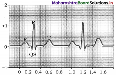
Answer:
‘T’ wave represents ventricular diastole.
Question 30.
Mention the role of pacemaker in human heart.
Answer:
Pacemaker can generate wave of contraction or cardiac impulse for rhythmic contraction of heart.
Question 31.
Which structure in the heart is called pacemaker?
Answer:
Sinuatrial node [S. A. node] in the heart wall is called a pacemaker.
Question 32.
What is electrocardiograph?
Answer:
The instrument which is used to record action potentials generated in the heart muscles is called an electrocardiograph or ECG machine.
Question 33.
What is angina pectoris?
Answer:
Angina pectoris is the pain in the chest caused due to reduction in blood supply to cardiac muscle caused due to narrowed and hardened coronary arteries.
Question 34.
What is pulse pressure?
Answer:
Difference between systolic and diastolic pressure is called pulse pressure which is normally 40 mm Hg.
Question 38.
What would happen if respiration takes place in one single step?
Answer:
If respiration takes place in one single step, then the chemical energy released at once during that step might result in a brief blast of light and heat and may lead to death of the cell. Hence respiration is a step-wise process.
Question 39.
Why do the veins have valves?
Answer:
The veins have valves at regular intervals to prevent backflow of blood as blood flows through veins with low pressure.
Question 40.
What is Bohr effect?
Answer:
Bohr effect is the shift of oxyhaemoglobin dissociation curve due to change in partial pressure of CO in blood.
Question 41.
What is Haldane effect?
Answer:
Decrease of pH of blood, due to increase in the number of H+ ions, HCO3 changes into H2O and CO2 by the presence of oxyhaemoglobin is called Haldane effect.
Give definitions of the following
Question 1.
Respiration
Answer:
It is a biochemical process of oxidation of organic compounds in an orderly manner for the liberation of chemical energy in the form of ATP.
Question 2.
Breathing
Answer:
It is a physical process by which gaseous exchange takes place between the atmosphere and the lungs. It involves inspiration and expiration.
Question 3.
Tidal Volume (TV)
Answer:
It is the volume of un¬ inspired or expired during normal breathing. It is 500 ml.
Question 4.
Inspiratory reserve volume (IRV)
Answer:
The maximum or the extra volume of air that is inspired during forced breathing in addition to TV (2000 to 3000 ml).
Question 5.
Expiratory reserve volume (ERV)
Answer:
The maximum volume of air that is expired during forced breathing after normal expiration. (1000 to 1100 ml).
Question 6.
Dead space (DS)
Answer:
The volume of air that is present in the respiratory tract (from nose to the terminal bronchioles), but not involved in gaseous exchange (150 ml).
Question 7.
Residual volume (RV)
Answer:
The volume of air that remains in the lungs and the dead space even after maximum expiration (1100 to 1200 ml).
Question 8.
Total lung capacity
Answer:
The maximum amount of air that the lungs can hold after a maximum forceful inspiration (5200 to 5900 ml).
Question 9.
Vital capacity (VC)
Answer:
The maximum amount of air that can be breathed out after of maximum inspiration. It is the sum total of TV, IRV and ERV and is 4100 to 4600 ml.
Question 10.
Oxygen dissociation curve
Answer:
The relationship between HbO2 saturation and oxygen tension (PPO2) is called oxygen dissociation curve.
Question 11.
Phosphorylation
Answer:
The process that involves trapping the heat energy in the form of high energy bond of ATP molecule is called phosphorylation.
Question 12.
Artificial ventilation
Answer:
It is the method of inducing breathing in a person when natural respiration has ceased or is faltering.
Question 13.
Ventilator
Answer:
A ventilator is a machine that supports breathing and is used during surgery, treatment for serious lung diseases or other conditions when normal breathing fails.
Question 14.
Cyclosis
Answer:
Cyclosis is the streaming movement of the cytoplasm shown by almost all living organisms. E.g. Paramoecium, Amoeba, etc.
Question 15.
Single circulation
Answer:
The movement of blood once through the heart during each circulation cycle is called single circulation.
Question 16.
Double circulation
Answer:
The movement of blood twice through the heart during one circulation cycle is called double circulation.
Question 17.
Erythropoiesis
Answer:
The process of formation of RBCs is called erythropoiesis.
Question 18.
Polycythemia
Answer:
The increase in the number of RBCs is called polycythemia.
Question 19.
Erythrocytopenia
Answer:
The decrease in the number of RBCs is called Erythrocytopenia.
Question 20.
Hematocrit
Answer:
The hematocrit is ratio of the volume of RBCs to total blood volume of blood.
Question 21.
Diapedesis
Answer:
Leucocytes perform amoeboid movement. Due to this kind of movement they can move out of the capillary walls. This is called diapedesis.
Question 22.
Leucocytosis
Answer:
Increase in the number of leucocytes or WBCs is called leucocytosis.
Question 23.
Leucopenia
Answer:
The decrease in the number of white blood cells is called leucopenia.
Question 24.
Leukaemia
Answer:
Pathological Increase in the number WBCs is called leukaemia or blood cancer.
Question 25.
Thrombocytopenia
Answer:
Decrease in the number of blood platelets is called thrombocytopenia.
Question 26.
Blood Coagulation
Answer:
Conversion of liquid blood into semisolid jelly is called blood coagulation or blood clotting.
Question 27.
Pericardium
Answer:
Double layered peritoneum that covers the heart from outside is called pericardium.
Question 28.
Pacemaker
Answer:
Pacemaker is the region that has power of generation of wave of contraction. In heart, sinoatrial node is called pacemaker.
Question 29.
Heartbeat
Answer:
The rhythmic contraction and relaxation of the heart is called heartbeat.
Question 30.
Pulse
Answer:
A pressure wave that travels through the arteries after each ventricular systole is called pulse.
Question 31.
Heart rate
Answer:
The rate with which the heart beats per minute is called the heart rate.
Question 32.
Stroke volume
Answer:
The amount of blood thrown out of the ventricles during one systole is called the stroke volume.
Question 33.
Cardiac output
Answer:
The amount of blood thrown out of the ventricles during one minute is called cardiac output.
Question 34.
Tachycardia
Answer:
Higher heart rate over 100 beats per minute is called tachycardia.
Question 35.
Bradycardia
Answer:
Lower heart rate which is lesser than 60 per minute is called bradycardia.
Question 36.
Myogenic
Answer:
When the initiation and further regulation of heartbeats take place in the muscles then such a heart is called myogenic.
Question 37.
Cardiac cycle
Answer:
Consecutive systole and diastole constitutes a single heartbeat or cardiac cycle.
![]()
Question 38.
Arterial blood pressure
Answer:
The pressure exerted by blood on the wall of artery is called arterial blood pressure.
Question 39.
Angiology
Answer:
Study of blood vessels is called angiology.
Question 40.
Angiography
Answer:
X-ray or imaging of the cardiac blood vessels to locate the position of blockages is called angiography.
Question 41.
Heart transplant
Answer:
Replacement of severely damaged heart by normal heart from brain- dead or recently dead donor is called heart transplant.
Question 42.
Silent Heart Attack
Answer:
Silent heart attack, also known as silent myocardial infarction, is a type of heart attack that lacks the general symptoms of classic heart attack like extreme chest pain, hypertension, shortness of breath, sweating and dizziness.
Question 43.
Electrocardiogram
Answer:
Graphical recording of electrical variations detected at the surface of body during their propagation through the wall of heart is electrocardiogram (ECG).
Question 44.
Lymph
Answer:
It is a fluid connective tissue with almost similar composition to the blood except RBCs, platelets and some proteins.
Give functions of the following
Question 1.
Epiglottis.
Answer:
The epiglottis prevents the entry of food into the trachea by closing the glottis temporarily.
Question 2.
Carbonic anhydrase.
Answer:
Carbonic anhydrase enzyme is found inside the RBCs only to accelerate the rate of formation of carbonic acid from CO2 and H2O.
Question 3.
Ventilators.
Answer:
Ventilators used in hospitals are part of life supporting system, which help in breathing by
- Pushing oxygen into the lungs
- Removing carbon dioxide from the lungs
Question 4.
Erythrocytes.
Answer:
Erythrocytes carry oxygen to all cells of the body from the lungs and bringing carbon dioxide from all the cells back to lungs.
Question 5.
Neutrophils.
Answer:
Neutrophils are responsible for destroying pathogens by the process of phagocytosis.
Question 6.
Thrombocytes/Platelets.
Answer:
Platelets secrete platelet factors which are essential in blood clotting. They also seal v the ruptured blood vessels by formation of platelet plug/thrombus. They secrete serotonin, a local vasoconstrictor.
Question 7.
Pericardial fluid.
Answer:
Pericardial fluid acts as a shock absorber and protects the heart from mechanical injuries. It also keeps the heart moist and acts as lubricant.
Question 8.
Heart walls.
Answer:
The epicardium and endocardium are protective in function whereas myocardium is responsible for contraction and relaxation of heart.
Question 9.
Valves in heart.
Answer:
Valves in the heart prevent the backflow of the blood at the time of systole and help in maintaining a unidirectional flow of blood.
Question 10.
Chordae tendinae.
Answer:
Chordae tendinae attach the bicuspid and tricuspid valves to the ventricular wall (papillary muscles) and regulate their opening and closing.
Question 11.
Semilunar valves.
Answer:
Semilunar valves prevent the backward flow of blood from pulmonary aorta and the aorta into the respective ventricles.
Question 12.
Sinoatrial node [SA] or Pacemaker.
Answer:
SA node acts as pacemaker of heart because it has the power of generating a new wave of contraction and making the pace of contraction.
Question 13.
Electrocardiogram (ECG).
Answer:
ECG helps to diagnose the abnormality in conducting pathway, enlargement of heart chambers, damage to cardiac muscles, reduced blood supply to cardiac muscles and causes of chest pain.
Question 14.
Blood.
Answer:
Functions of blood:
- Transport of oxygen and carbon dioxide
- Transport of food
- Transport of waste product
- Transport of hormones
- Maintenance of pH
- Water balance
- Transport of heat
- Defence against infection
- Temperature regulation
- Blood clotting/coagulation
- Helps in healing
Name the following
Question 1.
Name two animals in which moist skin acts as a respiratory surface.
Answer:
Earthworm, Frog
Question 2.
Name the respiratory organs in insects and fish.
Answer:
Insects – Tracheal tubes and spiracles
Fish – Internal gills
Question 3.
Name any two disorders of respiratory system.
Answer:
Asthma and pneumonia are the two disorders of respiratory system.
Question 4.
Name the structural and functional unit of lungs.
Answer:
Alveolus is the structural and functional unit of lungs.
Question 5.
Name the energy currency of cell.
Answer:
ATP is the energy currency of cell.
Question 6.
Name the site where actual exchange of O2 and CO2 takes place between air and blood in the body of man.
Answer:
Alveolus of lung.
Question 7.
Name any two respiratory centres required for regulation of breathing.
Answer:
Inspiratory centre, Expiratory centre, Pneumotaxic centre and Apneustic centre.
Question 8.
Name the muscles which move ribs up and down.
Answer:
External intercostal muscles.
Question 9.
Name two phyla where haemocoel is present.
Answer:
Phylum-Arthropoda and Phylum-Mollusca.
Question 10.
Name the animal-group which show single circulation.
Answer:
Fishes
Question 11.
Name the cells which produce thrombocytes.
Answer:
Megakaryocytes produce thrombocytes.
Question 12.
Name the process of formation of red blood corpuscles.
Answer:
Erythropoiesis
Question 13.
Name the space in which human heart is located.
Answer:
Mediastinum is the space in which human heart is located.
Question 14.
Name the types of lymphocytes depending upon functions.
Answer:
B-lymphocytes and T-lymphocytes.
Question 15.
Name the layers of peritoneum that surrounds the heart sequentially from outside to inside.
Answer:
Fibrous pericardium, parietal layer of serous pericardium and visceral layer of serous pericardium.
Question 16.
Name the connection between the pulmonary trunk and systemic aorta.
Answer:
Ligamentum arteriosum that represents remnant of ductus arteriosus of foetus.
Question 17.
Name the valve between left atrium and left ventricle and give its significance.
Answer:
Between left atrium and left ventricle is mitral or bicuspid valve which maintains the unidirectional flow of blood by preventing hs backflow.
Question 18.
Name the walls of an artery.
Answer:
Outer tunica externa, middle tunica media and inner tunica interna.
Question 19.
Name the instrument used to measure blood pressure.
Answer:
Sphygmomanometer is used to measure blood pressure.
Question 20.
Name the plasma proteins involved in the process of blood clotting.
Answer:
Prothrombin and fibrinogen.
Question 21.
Name the various components of conducting system of the heart.
Answer:
Conducting system of the heart consists of SA node, AV node, bundle of His and Purkinje fibers.
![]()
Question 22.
Name the neurotransmitters that decrease and increase the heart rate in human beings respectively.
Answer:
Acetylcholine decreases heart rate and adrenaline or epinephrine increases the heart rate in human.
Question 23.
Who discovered ECG?
Answer:
Willem Einthoven discovered ECG.
Distinguish between the following
Question 1.
Pharynx and Larynx.
Answer:
| Pharynx | Laryix |
| 1. Pharynx is a short, vertical tube. | 1. Larynx is a sound producing organ located at the end of pharynx. |
| 2. Mouth leads to the pharynx. | 2. Larynx leads to the oesophagus. |
| 3. Vocal cords are absent. | 3. Vocal cords are present. |
| 4. Pharynx does not increase in size at the time of puberty. | 4. Larynx increases in size at the time of puberty. |
| 5. Pharynx does not show Adam’s apple. | 5. Larynx shows Adam’s apple in adult males. |
Question 2.
Inspiration and Expiration.
Answer:
| Inspiration | Expiration |
| 1. Inspiration is an active process. | 1. Expiration is a passive process. |
| 2. During inspiration diaphragm contracts and becomes flattened. | 2. During expiration diaphragm relaxes and becomes dome shaped. |
| 3. During inspiration intercostal muscles contract. | 3. During expiration intercostal muscles relax. |
| 4. During inspiration ribs are pulled outwards and sternum is raised. | 4. During expiration ribs are pulled inwards and sternum is lowered. |
| 5. During inspiration the space in the thoracic cavity increases. | 5. During expiration the space in the thoracic cavity decreases. |
| 6. During inspiration pressure in the lungs decreases. | 6. During expiration pressure in the lungs increases. |
| 7. During inspiration the volume of the lungs increase. | 7. During expiration the volume of the lungs decreases. |
| 8. During inspiration air comes inside the body. | 8. During expiration air goes out of the body. |
Question 3.
External respiration and Internal respiration.
Answer:
| External respiration | Internal respiration |
| 1. The respiratory processes occurring in lungs is called external respiration. | 1. The respiratory processes that occur in tissues is called internal respiration. |
| 2. During external respiration O2 from the lungs enters into the lung capillaries by diffusion. | 2. During internal respiration O2 from the blood enters the tissue cells. |
| 3. During external respiration CO2 from the lung capillaries diffuse into the lungs. | 3. During internal respiration CO2 from the tissues enters into the blood. |
| 4. During external respiration exchange of gases takes place between the air and the lungs. | 4. During internal respiration exchange of gases take place between the blood and the tissue. |
| 5. Formation of oxyhaemoglobin takes place during external respiration. | 5. Oxyhaemoglobin dissociates into oxygen and haemoglobin during internal respiration. |
| 6. During external respiration CO2 is released. | 6. During internal respiration carbamino haemoglobin is formed which is carried to the lungs. |
Question 4.
Transport of O2 and Transport of CO2.
Answer:
| Transport of O2 | Transport of CO2 |
| 1. Transport of O2 takes place from lungs to the tissues and cells. | 1. Transport of CO2 takes place from tissues and cells to the lungs. |
| 2. Oxygen is carried as oxyhaemoglobin to the tissues with the help of RBCs. | 2. Carbon dioxide is carried as carbaminohaemoglobin from the tissues with the help of plasma and RBCs. |
| 3. Oxygen does not form oxides or other products during its transport. | 3. CO2 forms bicarbonates with sodium and potassium during its transport. |
| 4. O2 does not form acids during its transport. | 4. CO2 dissolves in water to form carbonic acid. |
Question 5.
Vital Capacity of Lung and Total Lung Capacity.
Answer:
| Vital Capacity of Lung | Total Lung Capacity |
| 1. It is the maximum amount of air a person can expire and inspire to their maximum extent. | 1. It is the maximum amount of air that the lungs can hold after a maximum forceful inspiration. |
| 2. It is the sum total of inspiratory reserve volume, tidal volume and expiratory reserve volume. | 2. It is the sum total of vital capacity and residual volume. |
| 3. It ranges from 4100 to 4600 ml. | 3. It ranges from 5200 to 5800 ml. |
Question 6.
Inspiratory Reserve Volume (IRV) and Expiratory Reserve Volume (ERV).
Answer:
| Inspiratory Reserve Volume (IRV) | Expiratory Reserve Volume (ERV). |
| 1. It is the maximum volume of air, or the extra volume of air, that is inspired during forced breathing. | 1. It is the maximum volume of air that is expired during forced breathing. |
| 2. Its value is 2000/3000 ml. | 2. Its value is 1000/1100 ml. |
Question 7.
T. S. of artery and T.S. of vein.
Answer:
| T. S. of artery | T.S. of vein |
| 1. Histologically in transverse section of artery there are three walls, tunica externa, tunica media and tunica interna or endothelium. | 1. Histologically in transverse section of vein there are three walls, tunica externa, tunica media and tunica interna or endothelium. |
| 2. Tunica media is thick and muscular. | 2. Tunica media is thinner as compared to artery. |
| 3. Lumen of artery is narrow. | 3. Lumen of vein is broad. |
Question 8.
Erythrocytes and Leucocytes.
Answer:
| Erythrocytes | Leucocytes |
| 1. Erythrocytes have a definite shape which is elliptical or oval. | 1. Leucocytes do not have definite shape as they are amoeboid. |
| 2. They are enucleated. | 2. They are nucleated. |
| 3. Erythrocytes contain haemoglobin and hence appear red. | 3. Leucocytes are devoid of any respiratory pigment and hence appear colourless. |
| 4. The normal erythrocyte count is 4.3 to 5.8 million per cubic mm of blood. | 4. The normal leucocyte count is 4000 to 11000 per cubic mm of blood. |
| 5. The life span of erythrocytes is 100 to 120 days. | 5. The life span of leucocytes is 3 to 4 days. |
| 6. The diameter of erythrocytes is 7.2 m and thickness is about 2 to 2.2 m. | 6. The size of leucocytes varies with its subtypes and is of average size of 8 to 15 m. |
| 7. Erythrocytes are formed by the process of erythropoiesis in red bone marrow. | 7. Leucocytes are formed by the process of leucopoiesis in bone marrow, tonsils, lymph nodes, spleen, thymus, etc. |
| 8. Erythrocytes transport the respiratory gases. | 8. Leucocytes help in the formation of antibodies besides fighting against foreign antigens by phagocytic activity. |
Question 9.
Eosinophils and Basophils.
Answer:
| Eosinophils | Basophils |
| 1. Cytoplasmic granules present in eosinophils are stained with acidic stains. | 1. Cytoplasmic granules present in basophils are stained with basic stains. |
| 2. Nucleus is bilobed. | 2. Nucleus is twisted. |
| 3. Eosinophils constitute 3% of total WBCs. | 3. Basophils constitute 0.5% of total WBCs. |
Question 10.
Neutrophils and Eosionophils.
Answer:
| Neutrophils | Eosinophils |
| 1. Cytoplasmic granules present in neutrophils are stained with neutral stains. | 1. Cytoplasmic granules present in eosinophils are stained with acidic stains. |
| 2. Nucleus is three to five lobes showing polymorphic form. | 2. Nucleus is bilobed. |
| 3. Neutrophils constitute 62% of total WBCs. | 3. Eosinophils constitute 3% of total WBCs. |
Question 11.
Lymphocytes and Monocytes.
Answer:
| Lymphocytes | Monocytes |
| 1. Large round nucleus but size of the cell is smaller. | 1. Large kidney shaped nucleus and largest size among WBCs. |
| 2. Lymphocytes form 25-33% of WBCs. | 2. Monocytes form 3-9% of WBCs. |
Question 12.
Granulocytes and Agranulocytes.
Answer:
| Granulocytes | Agranulocytes |
| 1. WBCs with granular cytoplasm are called granulocytes. Thus, cytoplasmic granules are present. | 1. WBCs with agranular cytoplasm are called agranulocytes. Thus, cytoplasmic granules are absent. |
| 2. Nuclei of granulocytes are variously lobed. | 2. Nuclei of agranulocytes are not lobed. |
Question 13.
Single circulation and Double circulation.
Answer:
| Single circulation | Double circulation |
| 1. Blood flows only once through the heart in a complete cycle. | 1. Blood flows twice through the heart during one complete circulation. Systemic – to and fro ‘ from heart to body and pulmonary – to and fro from heart to lungs. |
| 2. Heart pumps deoxygenated blood only. | 2. Heart pumps both deoxygenated and oxygenated blood to lungs and body respectively. |
| 3. Blood is oxygenated in gills. | 3. Blood is oxygenated in lungs. |
| 4. Occurs only in fishes. | 4. Occurs in amphibians, reptiles, birds and mammals. |
Question 14.
Systolic blood circulation and Diastolic blood circulation.
Answer:
| Systolic blood circulation | Diastolic blood circulation |
| 1. Blood is passed from right ventricle to lungs by pulmonary artery during systolic circulation. Similarly, from left ventricle the oxygenated blood is given to the entire body through systemic aorta during systolic circulation. | 1. Blood is passed to left atrium from lungs by pulmonary veins during diastolic circulation. Similarly, deoxygenated blood from entire body is brought back to heart through vena cava during diastolic circulation. |
| 2. Systolic blood circulation is under maximum pressure as heart is forcing the blood to come out of heart. | 2. Diastolic blood circulation is under minimum blood pressure as heart is relaxed during diastole. |
Question 15.
Atria and Ventricles.
Answer:
| Atria | Ventricles |
| 1. Atria are upper chambers of the heart. | 1. Ventricles are lower chambers of the heart. |
| 2. Atria are thin walled. | 2. Ventricles are thick walled. |
| 3. Atria are receiving chambers. | 3. Ventricles are distributing chambers. |
| 4. Interatrial septum divides the two auricles (atria). | 4. Interventricular septum divides the two ventricles. |
| 5. Right atrium is larger in size than left atrium. | 5. Left ventricle is larger in size than the right ventricle. |
Question 16.
S.A. Node and A.V. Node.
Answer:
| S.A. Node | A.V. Node |
| 1. Sinoatrial node is present in the right ventricle near the opening near the opening of the superior vena cava. | 1. Atrioventricular node is present in the right ventricle near the opening of the coronary sinus. |
| 2. S.A. node is the pacemaker of the heart and it starts atrial systole. | 2. A.V. node starts ventricular systole through bundles of His and Purkinje’s fibre system. |
Question 17.
Pulmonary circulation and Systemic circulation.
Answer:
| Pulmonary circulation | Systemic circulation |
| 1. The course of blood from the right ventricle to the left atrium of the heart through the lungs is called pulmonary circulation. | 1. The course of blood from the left ventricle to the right atrium of the heart through the body is called systemic circulation. |
| 2. Pulmonary circulation is mainly for sending the blood for oxygenation in the lungs from the heart and bringing it back to the heart after oxygenation. | 2. Systemic circulation is for sending the deoxygenated blood from the body to the heart and sending oxygenated blood from the heart to the body. |
Question 18.
Atrio ventricular valves and Semilunar valves.
Answer:
| Atrio ventricular valves | Semilunar valves |
| 1. Atrio ventricular valves Eire present between the atria and ventricles. On the right side there is tricuspid valve whereas on the left side there is bicuspid valve. | 1. Semilunar valves are present at the opening of pulmonary artery and systemic aorta. |
| 2. Atrio ventricular valves prevent the back flow of blood from ventricles to atria at the time of systole. | 2. Semilunar valves prevent the back flow of blood from pulmonary artery and systemic aorta back to the heart. |
Question 19.
Hypertension and Hypotension.
Answer:
| Hypertension | Hypotension |
| 1. Blood pressure values more than 140 mm Hg SP and 90 mm HG DP is called hypertension. | 1. Blood pressure values less them 120 mm Hg SP and 70 mm HG DP is called hypotension. |
| 2. Excessive hypertension can result into lethal complications such as stroke or paralysis. | 2. Hypotension may not be lethal if immediate measures are taken to raise the blood pressure. |
Give reasons
Question 1.
ATP is called energy currency of the cell.
Answer:
- During cellular respiration, the oxidation of food (glucose) takes place.
- This happens in the mitochondria using the oxygen present in the blood.
- ATP molecules are formed during this oxidation.
- ATP is used for various vital body processes and also for maintaining the body temperature to constancy.
- Since energy is stored in the form of ATP it is called an energy currency of the cell.
Question 2.
The vestibule of nasal chamber has fine hair.
Answer:
- Vestibule is the anterior most part of the nasal chamber.
- The hairs present in this region trap the dust particles and prevent them from entering into the interiors of the respiratory passage.
- Therefore, the vestibule of nasal chambers has fine hair.
Question 3.
Glottis is guarded by a flap called epiglottis.
Answer:
- The oesophagus and trachea lie side by side.
- There is possibility that food particles may enter respiratory passage at the time of gulping.
- However, the epiglottis prevents the entry of food into the respiratory passage by closing it temporarily.
- Thus, for preventing the entry of food particles into respiratory passage, the glottis is guarded by a flap called epiglottis.
![]()
Question 4.
Alveoli are very flexible.
Answer:
- Alveoli are made up of collagen and elastin fibres.
- They are very thin (0.0001 mm) and lined by non-ciliated squamous epithelium.
- All the above structural components make the alveoli very flexible.
Question 5.
Expiration is called a passive process.
Answer:
- During expiration, intercostal muscles relax. This results in pulling the ribs inwards.
- Diaphragm also relaxes and returns to its normal dome shape.
- The collective contraction of ribs and diaphragm results in the reduction of
thoracic cavity and hence automatically air is pushed out of the lungs. - Since the pressure on the lungs increase rushing the air to outside, expiration is called a passive process.
Question 6.
Pericardium acts as a defence wall for the heart.
Answer:
- Pericardium protects the heart. It is double layered peritoneum, having outer fibrous and inner serous pericardium layers.
- Fibrous pericardium being tough gives protection to the heart.
- Serous pericardium has two layers, parietal and visceral layer or epicardium.
- In between these two layers, there is pericardial fluid, which helps to absorb shocks and provide nourishment.
- In this way pericardium acts as a defence wall.
Question 7.
Valves are present in veins.
Answer:
- Veins carry blood to the heart.
- At that time the backward flow of the blood should be prevented.
- Therefore, valves are present in veins.
Question 8.
Atria are thin walled than ventricles.
Answer:
- Atria are receiving chambers, while ventricles are distributing chambers.
- The blood is driven out from ventricles.
- Ventricles are therefore, strong and with thicker walls.
- Atria are thin walled as compared to ventricles.
Question 9.
Heart is called a pump.
Answer:
- The heart acts as a pumping organ. It shows continuous pumping action.
- The rhythmic contraction or systole and relaxation or diastole of heart forms one heartbeat.
- Such heartbeats occur about 72 times per minute.
- The heart efficiently pumps about 5 litres of blood per minute. Therefore, the heart is called a pump.
Question 10.
In normal human heart, there is no mixing of oxygenated and deoxygenated blood.
Answer:
- In normal human heart, there is completely formed atrioventricular septum.
- This septum keeps the deoxygenated and oxygenated blood separate.
- Hence there is no mixing of the two types of blood.
Question 11.
Blood pressure is inversely related to the elasticity of the blood vessels.
Answer:
- When the blood gushes through the blood vessels, the walls of blood vessels -can expand a little due to their elasticity.
- But as the age advances, the elasticity is reduced and then the blood vessels do not expand.
- Hence the flowing blood gets more resistance and the blood pressure can rise.
- Lesser the elasticity more will be the blood pressure, whereas more the elasticity of the vessel wall, then the pressure will not rise.
- In this way, the blood pressure is inversely related to the elasticity of the blood vessels.
Write short notes
Question 1.
Chloride shift or Hamburger’s phenomenon.
Answer:
- About 70% of CO2 is transported in the form of sodium bicarbonates/potassium bicarbonates from tissue cells to lungs.
- In the RBCs, CO2 combines with water in the presence of a Zn containing enzyme, carbonic anhydrase to form carbonic acid. This action is rapid in RBCs as compared to that in the plasma.
- Carbonic acid being unstable, immediately dissociates into HCO3– and H+in the presence of same enzyme, leading to large accumulation of HCO3– inside the RBCs. It thus moves out of RBCs. This can bring about imbalance of the charge inside the RBCs.
- To maintain the ionic balance between the RBCs and the plasma, Cl– diffuses into the RBCs. This movement of chloride ions is known as chloride shift or Hamburger’s phenomenon.
- HCO3– that comes in the plasma joins to Na+/K+ forming NaHCO3/KHCO3 which can maintain pH of blood. The remaining H+ ions in the RBCs are buffered by haemoglobin by the formation of oxyhaemoglobin.
- At the level of lungs, due to the low partial pressure of carbon dioxide of the alveolar air, hydrogen ion and bicarbonate ions combine to form carbonic acid and under the influence of carbonic anhydrase again yields carbon dioxide and water.
Question 2.
Regulation of breathing.
Answer:
(1) Respiration is under dual control, i.e. nervous and chemical. Normal breathing is an involuntary process. Steady state of respiration is controlled by neurons located in the pons and medulla and are known as the respiratory centres. They regulate the rate and depth of breathing.
(2) These centres are divided into three groups : dorsal group of neurons in the medulla (inspiratory centre), ventro-lateral group of neurons in medulla (inspiratory and expiratory centre) and pneumotaxic centre located in the pons and apneustic centre which is antagonistic in action to pneumotaxic centre.
(3) During inspiration, when the lungs expand to a critical point, the stretch receptors are stimulated and impulses are sent along the vagus nerves to the expiratory centre. It then sends out inhibitory impulses to the inspiratory centre.
(4) The inspiratory muscles relax and expiration follows. As the air leaves but, the lungs are deflated and the stretch receptors are no longer stimulated. Thus, the inspiratory centre is no longer inhibited and a new respiration begins. These events are called the Hering – Breuer reflex. The Hering – Breuer reflex controls the depth and rhythm of respiration. It also prevents the lungs from inflating to the point of bursting.
(5) The respiratory centre has connections with the cerebral cortex that means we can voluntarily change our pattern of breathing. Voluntary control is protective because it enables us to prevent water or irritating gases from entering the lungs.
Question 3.
Carbon monoxide poisoning.
Answer:
- Carbon monoxide poisoning is caused when carbon monoxide is combined with haemoglobin.
- Haemoglobin is said to have 250 times more affinity for carbon monoxide than that for the oxygen.
- Therefore, haemoglobin with carbon monoxide forms a stable compound, the carboxyhemoglobin.
- Due to the formation of carboxyhaemoglobin, the haemoglobin no longer carries oxygen to the cells and tissues. Tissues then suffer from oxygen starvation. This leads to asphyxiation and in extreme cases it leads to death.
- Carbon monoxide poisoning occurs in closed rooms with incompletely burning substances such as stove burners or furnaces and garages having running automobile engines.
- Person suffering from carbon monoxide poisoning has to be administered with oxygen-carbon dioxide mixture, so that high levels of CO2 makes carbon monoxide dissociated from haemoglobin.
Question 4.
Artificial ventilation.
Answer:
(1) Artificial ventilation is the artificial respiration. It is the method of inducing breathing in a person when natural respiration has ceased or is faltering. If used properly and quickly, it can prevent death due to drowning, choking, suffocation, electric shock, etc.
(2) The process involves two main steps:
a. Establishing and maintaining an open air passage from the upper respiratory tract to the lungs.
b. Force inspiration and expiration as in mouth to mouth respiration or by mechanical means like ventilator.
(3) A ventilator is a machine that supports breathing and is used during surgery, treatment for serious lung diseases or other conditions when normal breathing fails.
Question 5.
Erythrocytes.
Answer:
- Erythrocytes or red blood corpuscles. They are circular, biconcave, enucleated cells.
- The RBC size : 7 pm in diameter and 2.5 pm in thickness.
- The RBC count : 5.1 to 5.8 million RBCs/ cu mm of blood in an adult male and 4.3 to 5.2 million/cu mm in an adult female.
- The average life span of RBC : 120 days.
- RBCs are formed by the process of erythropoiesis. In foetus, RBC formation takes place in liver and spleen whereas in adults it occurs in red bone marrow.
- The old and worn out RBCs are destroyed in liver and spleen.
- Polycythemia is an increase in number of RBCs while erythrocytopenia is decrease in their (RBCs) number.
Functions of RBCs:
- Transport of oxygen from lungs to tissues and carbon dioxide from tissues to lungs with the help of haemoglobin.
- Maintenance of blood pH as haemoglobin acts as a buffer.
- Maintenance of the viscosity of blood.
Question 6.
Heartbeat.
Answer:
- The rhythmic contraction and relaxation of the heart is called heartbeat.
- Each heartbeat includes one systole and one diastole. During systole the heart contracts and during diastole it relaxes.
- The rate with which the heart beats is called heart rate. The normal heart rate is 72 beats per minute.
- Tachycardia means faster heart rate of about more than 100 beats per minute.
- Bradycardia means slower heart rate that is below 60 beats per minute.
Question 7.
Pulse.
Answer:
- A pressure wave that travels through the arteries after each ventricular systole is called a pulse.
- The pulse can be felt in any artery that lies near the surface of the body.
- The radial artery at the wrist is most commonly used to feel the pulse.
- The pulse rate per minute indicates the heart rate. Since each heartbeat generates one pulse in the arteries, the pulse rate is same as that of heart rate, i.e. 72 times per minute.
![]()
Question 8.
Peacemaker.
Answer:
- Pacemaker is the region in tile heart which initiates the beating.
- The natural pacemaker of the heart is sinoatrial node (SA node).
- The pacemaker is autorhythmic, it is able to repeatedly and rhythmically generate impulses.
- SA node is responsible for initiation of cardiac excitation. Therefore, it is called a pacemaker.
Question 9.
Blood pressure.
Answer:
- Blood pressure is the pressure exerted by the flowing blood on the walls of arteries.
- Blood pressure described in two terms viz. systolic blood pressure and diastolic blood pressure. Systolic blood pressure is the maximum pressure of blood when heart undergoes ventricular systole. It is responsible for flow of blood in the arteries. Normal systolic pressure is 120 mm Hg.
- Diastolic blood pressure is the minimum pressure of blood when heart undergoes diastole. Normal diastolic pressure is 80 mm Hg.
- Blood pressure is represented as 120/80 mm Hg for a normal human being.
Question 10.
Hypertension.
Answer:
- In a normal healthy person the blood pressure values are 120 mm Hg (systolic)/ 80 mm Hg (diastolic).
- When the blood pressure is persistently more than 140 mm Hg systolic pressure and 90 mm Hg diastolic arterial blood pressure then it is said to be hypertension or high blood pressure.
- Excessively high blood pressure is very dangerous as high blood pressure of about 220/120 mm Hg may cause rupturing of blood vessels.
- Rupture of eye blood vessels can lead to blindness.
- If blood vessels of kidney are affected then nephritis is caused.
- Hemorrhage occurring in the brain can lead to stroke or paralysis. Therefore, hypertension is commonly called silent killer. It may be present for years with no distinct symptoms.
- The factors causing hypertension are arteriosclerosis (reduction of elasticity of blood vessels), atherosclerosis (deposition, of cholesterol inside the blood vessels wall), obesity, physical or emotional stress, alcoholism, smoking, cholesterol rich diet, increased secretion of renin, epinephrine or aldosterone, etc.
Question 11.
Coronary artery disease (CAD).
Answer:
- Coronary artery disease is a condition caused due to problems like atherosclerosis.
- In this disease, coronary arteries are narrowed due to deposition of fatty substances.
- Due to this the blood flow to the heart is reduced.
- In coronary heart disease, the heart muscle is damaged because of an inadequate amount of blood due to obstruction of its blood supply.
- The symptoms of CAD depend upon the degree of obstruction.
- Symptoms are mild chest pain or angina pectoris.
- In severe cases it results in heart attack known as myocardial infarction.
Question 12.
Angina pectoris.
Answer:
- Angina pectoris is the pain in the chest. It results from a reduction in blood supply to cardiac muscle due to narrowed and hardened coronary arteries.
- Atherosclerosis and arteriosclerosis can cause this problem. Basically, the coronary arteries are affected during angina pectoris.
- It causes heaviness and severe pain in the chest. The pain can also be felt at the neck, lower jaw, left arm and left shoulder.
- Angina pectoris often occurs during exertion, when the heart demands more oxygen and narrowed blood vessels cannot supply. It disappears with rest.
Question 13.
Heart failure.
Answer:
- Heart failure is caused due to progressive weakening of the heart muscle. This results in the failure of the heart to pump the blood effectively.
- Hypertension increase the after load on the heart leading to significant enlargement of the heart.
- This finally results in heart failure.
- Factors responsible for heart failure are advanced age, malnutrition, chronic infections, toxins, severe anaemia or hyperthyroidism, etc.
- Any problem leading to degeneration of heart muscle, may result in heart failure.
Question 14.
Atherosclerosis.
Answer:
- Atherosclerosis is the deposition of fatty substances and cholesterol on the inner lining of eateries.
- This deposition results in the formation of atherosclerotic plaque.
- It results in the decrease of the lumen of the blood vessels causing increasing resistance for the blood to flow which in turn results in the hypertension.
- Atherosclerosis of the coronary arteries results in decrease in the blood flow to the heart muscles.
- Due to such condition, coronary heart disease is caused.
Question 15.
ECG.
Answer:
- Electrocardiogram or ECG is the graphic v record of electrical variations produced by the heart during one heartbeat or cardiac cycle.
- ECG is taken with the help of an instrument called electrocardiograph or ECG machine. Electrocardiograph records the action potentials generated by the heart muscles.
- The electrical activity of heart is represented in the form of a graph plotted with time on X-axis against voltage displacement on Y-axis.
- A normal ECG is a graph having series of ridges and furrows. There are waves such as P-wave, QRS complex and T-wave.
- P-wave is a small upwards wave representing impulse generated by SA node. P-wave is caused by atrial depolarization that results in atrial contraction.
- QRS-complex wave begins as a downward deflection, continues as a large upright triangular wave and ends’ as a downward wave.
- QRS-complex wave represents spreading of impulse from SA node to AV node, then to bundle of His and Purkinje fibres. It causes ventricular depolarization resulting in ventricular contraction.
- T-wave is a broad upward wave which represents ventricular repolarization resulting in ventricular relaxation.
- Functions of ECG are mainly for diagnosis and also for prognosis. It is useful to detect abnormal functioning of heart as in coronary artery diseases, heart block, angina pectoris, tachycardia, ischemic heart disease, myocardial infarction, cardiac arrest, etc.
Question 16.
Angiography.
Answer:
- Angiography is an X-ray imaging of the cardiac blood vessels to locate the position of blockages.
- Depending upon the degree of blockage, remedial procedures like angioplasty or by¬pass surgery are performed.
- In angioplasty a stent is inserted at the site of blockage to restore the blood supply while in by-pass surgery, the atherosclerotic region is by-passed with part of vein or artery taken from any other suitable part of the body, like hands or legs.
Question 17.
Silent Heart Attack or silent myocardial infarction.
Answer:
- Silent heart attack is a type of heart attack that lacks the general symptoms of classic heart attack like extreme chest pain, hypertension, shortness of breath, sweating and dizziness.
- Symptoms of silent heart attack are so mild that a person often confuses it for regular « discomfort and thereby ignores it.
- Men are more affected by silent heart attack than women.
Question 18.
Heart Transplant.
Answer:
- Heart transplant is the replacement of severely damaged heart by normal heart from brain-dead or recently dead donor,
- Heart transplant is necessary in case of patients with end-stage heart disease and severe coronary arterial disease.
Short Answer Questions
Question 1.
What is meant by respiration? How is it useful in the production of energy?
Answer:
- Respiration is the biochemical process in which organic compound such as glucose are oxidized to liberate chemical energy.
- During respiration energy is released in gradual and step wise process. The released energy is in the form of bonds of ATP (Adenosine Tri Phosphate) molecules are shown below:
C6H12O6 + 6O2 → 6CO2 + 6H2O + 38 ATP - ATP is the biologically useful energy. ATP drives most of the life process.
- When cell requires the energy, ATP is hydrolyzed and is converted into ADP with subsequent release of energy.
- The respiratory system, blood and the body cells play an important role in the process of respiration.
Question 2.
How does exchange of gases take place at the alveolar level?
Answer:
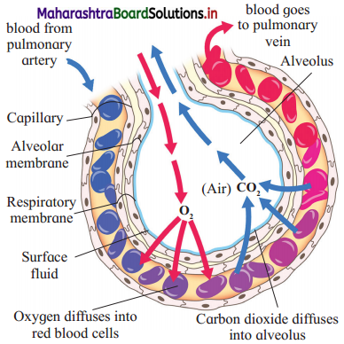
1. Exchange of gases between the alveolar air and the blood is known as external respiration.
2. Simple squamous epithelial layer of alveolus is intimately associated with a similar layer lining the capillary wall. Both of these layers are thin walled and together they make up the respiratory membrane through which gaseous exchange occurs between the alveolar air and the blood.
3. Diffusion of gases will take place from an area of higher partial pressure to an area of lower partial pressure until the partial pressure in the two regions reaches equilibrium.
4. The partial pressure of carbon dioxide of blood entering the pulmonary capillaries is 45 mmHg while partial pressure of carbon dioxide in alveolar air is 40 mmHg. Due to this difference, carbon dioxide diffuses from the capillaries into the alveolus.
5. Similarly, partial pressure of oxygen of blood in pulmonary capillaries is 40 mmHg while in alveolar blood it is 104 mmHg. Due to this difference oxygen diffuses from alveoli to the capillaries.
Question 3.
What is the role of haemoglobin in the transport of oxygen in the blood?
Answer:
- Haemoglobin is a respiratory pigment present in cytoplasm of RBCs. About 97% of oxygen is transported by these haemoglobin molecules from lungs to tissues.
- Haemoglobin has a high affinity for Oa and combines with it to form oxyhaemoglobin. One molecule of Hb has four FeT, each of which can pick up a molecule of oxygen (O2). Hb + 4O2 → Hb (4O2)
- Oxyhaemoglobin is transported from lungs to the tissues where it readily dissociates to release O2.
Hb (4O2) → Hb + 4O2 - In the alveoli where PPOa is high and PPCO2 is low, oxygen binds with haemoglobin, but in tissues, where PPO2 is lower and PPCO2 is high, Oxyhaemoglobin dissociates and releases O2 for diffusion into the tissue cells.
Question 4.
What is blood? What is the normal quantity of blood in an adult human being?
Answer:
- Blood is the fluid connective tissue that circulates in the body.
- Blood is derived from mesoderm.
- It is bright red, slightly alkaline fluid having pH about 7.4. It is salty, viscous fluid heavier than water.
- The average sized adult has about 5 litres of blood in his/her body which constitutes about 8% of the total body weight.
![]()
Question 5.
Describe the structure and the function of thrombocytes.
Answer:
- Thrombocytes or platelets are non- nucleated, round and biconvex blood corpuscles.
- They are smallest corpuscles measuring about 2.5 to 5 mm in diameter with a count of about 2.5 lakhs/cu mm of blood.
- Their life span is about 5 to 10 days.
- Thrombocytes are formed from megakaryocytes of bone marrow. They break from these cells as fragments during the process of thrombopoiesis.
- Thrombocytosis is the increase in platelet count while thrombocytopenia is decrease in platelet count.
- Thrombocytes possess thromboplastin which helps in clotting of blood.
- Therefore, at the site of injury platelets aggregate and form a platelet plug. Here they release thromboplastin due to which further blood clotting reactions take place.
Question 6.
Describe the structure of the heart wall.
Answer:
- The heart wall is composed of three layers, viz. outer epicardium, middle myocardium and inner endocardium.
- Epicardium is composed of single layer of mesothelium having flat epithelial cells.
- Myocardium is composed of cardiac muscle fibres. These muscle fibres perform the function of systole and diastole by showing contraction and relaxation of muscle wall of the heart.
- Endocardium is composed of single layer of flat epithelial cells called endothelium.
Question 7.
Name the two heart sounds. How and when are they produced?
Answer:
- In one normal heartbeat the heart sounds like lubb and dup are produced once each.
- The rhythmic contraction (Systole) and relaxation (diastole) forms are heartbeat. The heart sounds are due to closure of valves.
- Lubb sound is produced during ventricular systole when the cuspid valves close both the atrioventricular apertures preventing blood flow into atria.
- Dub sound is produced during ventricular diastole when semilunar valves are closed, preventing backflow of blood from pulmonary trunk and systemic aorta into ventricles.
Question 8.
What is double circulation? What is its significance?
Answer:
(1) Double circulation : Movement of blood twice through the heart during one circulation cycle is called double circulation. Body → heart → lungs → heart → body is the course of double circulation.
(2) Significance of double circulation:
a. Double circulation is more effective type of circulation in which oxygenated and deoxygenated type of blood do not intermix.
b. The systemic circulation i.e. from body to heart and back to body while the pulmonary circulation i.e. from heart to lungs and back to heart circulate the blood uniformly.
(3) Coronary and hepatic portal circulation is also achieved due to double circulation.
Question 9.
Describe pulmonary and systemic circulation.
Answer:
- In human beings, there is double circulation because blood passes twice through the heart during one cardiac cycle.
- The blood follows two routes viz. pulmonary and systemic.
- Pulmonary circulation is circulation between heart and lungs. Systemic circulation is the circulation between the heart and body organs (except lungs).
- During pulmonary circulation, the blood passes from the right ventricle to the left atrium of the heart through lungs.
- The right ventricle pumps deoxygenated blood into the pulmonary trunk which carries it to lungs for oxygenation. The oxygenated blood from the lungs is brought to left atrium by two pairs of pulmonary veins.
- During systemic circulation, the blood from the left ventricle passes to the right atrium of heart through body organs.
- The left ventricle pumps oxygenated blood into the systemic aorta which carries it to all body organs except lungs. The deoxygenated blood from the body organs is brought to right atrium by superior and inferior venae cavae.
Question 10.
How is cardiac activity regulated?
Answer:
- Normal activities of the heart are auto regulated. The specialized muscles help in this regulation.
- The heart is said to be myogenic due to this ability.
- In the medulla oblongata of brain, there is cardiovascular centre,
- From this centre, sympathetic and parasympathetic nerves innervate the sinoatrial node.
- Sympathetic nerves secrete adrenaline and it stimulates and increases the heartbeat.
- Parasympathetic nerves secrete acetylcholine and it decreases the heart rate.
- Rate of heartbeat is controlled in response to inputs from various receptors like proprio-receptors.
- These receptors monitor the position of limbs and muscles. There are chemoreceptors which monitor chemical change in blood and baroreceptors that monitor the stretching of main arteries and veins.
Chemical control on heart rate:
- Hypoxia, acidosis, alkalosis cause decrease in cardiac activity.
- Hormones like epinephrine and nor epinephrine enhance the cardiac activity.
- Elevated blood level of K+ and Na+ decreases the cardiac activity.
Question 11.
What are the main features of respiratory surface?
Answer:
The respiratory surface, for the efficient gaseous exchange should have the following features:
- It should have a large surface area.
- It should be thin, highly vascular and permeable to allow exchange of gases.
- It should be moist.
Question 12.
What is the co-relationship between activeness of organism and complexity of transport system?
Answer:
- As the size of an organism increases, its surface area to volume ratio decreases. This means it has relatively less surface area available for substances to diffuse through.
- Large multicellular organisms therefore cannot rely on diffusion alone to supply their cells with Substances such as food and oxygen and to remove waste products. Large multicellular organisms require specialized transport system.
- In short for the organisms to become active, they must be having complex transport system to bring about their vital functions rapidly.
Question 13.
What are the granules in granulocytes?
Answer:
- Neutrophils : Granules . of neutrophils contain cationic proteins and other proteins that are used to kill bacteria, some enzymes to breakdown bacterial proteins, lysozymes to breakdown bacterial cell wall. etc.
- Eosinophils : Granules of eosinophil contains a unique toxic basic protein receptors that bind to IgE used to help in killing parasites.
- Basophils : Granules of basophils contain abundant histamine, heparin and platelet activating factor.
Question 14.
What is haemoglobin count in normal human beings? What is the function of haemoglobin?
Answer:
- The normal haemoglobin in adult male is 13-18 mg/100 ml of blood.
- In a normal adult female, it is about 11.5-16.5 mg/100 ml of blood.
- In anaemic individuals there is lesser amount of haemoglobin.
- Functions of haemoglobin is to transport oxygen from lungs to tissues and carbon dioxide from tissues of lungs.
- Haemoglobin acts as a buffer and maintains the blood pH.
Question 15.
Why has the heart-recipient to rely upon life-time supply of immunosuppressants?
Answer:
Person who has undergone heart transplant needs lifetime supply of immunosuppressants because in these persons organ rejection is a constant threat. Keeping away the immune system from attacking the transplanted organ requires constant supply of immunosuppressant drugs. These drugs help prevent immune system from attacking (“rejecting”) the donor organ. Typically, these drugs are taken for the life-time for maintaining transplanted organ.
Question 16.
Why is it difficult to hold one’s breath beyond a limit?
Answer:
- It is difficult to hold one’s breath beyond a limit because the pressure of oxygen and carbon dioxide in blood changes as one holds his breath.
- When the breath is held beyond a limit, the urge to breath becomes irresistible.
- When the breath is held forever, body becomes starved of oxygen and person may fall unconscious and the instinct to breath would take over.
Question 17.
Why and when do the leucocytes perform diapedesis?
Answer:
- Diapedesis is the movement of leucocytes through the wall of blood capillaries into the tissue space.
- Leucocytes perform diapedesis as an important part of their reaction to tissue injury or infection.
- This process forms the part of the innate immune response, involving recruitment of non-specific leucocytes.
- Monocytes also use this process during their development into macrophages.
- Diapedesis helps leucocytes to perform their functions like phagocytosis, production of antibodies, secretion of inflammatory response chemicals, etc.
Question 18.
Why are obese persons prone to hypertension?
Answer:
(1) Being overweight or obese is a major cause of hypertension, accounting for 65% to 75% of the risk for human primary hypertension.
(2) Following factors play an important role in initiating obesity hypertension:
- Physical compression of the kidneys by fat.
- Activation of the renin-angiotensin – aldosterone system
- Increased sympathetic nervous system activity.
- Obesity means more body-surface area. In order to supply blood to these parts. Heart and blood vessels work more resulting into hypertensions.
(3) Blood pressure rises as body weight increases and therefore obese persons are prone to hypertension.
Question 19.
Why does the transplanted heart beats at higher rate than normal?
Answer:
- The transplanted heart beats at higher rate than normal (about 100 to 110 beats per minute) because the nerves leading to the heart are cut during the operation. These nerves stimulate the pacemaker i.e. Sinoatrial node.
- The new heart also responds more slowly to exercise and does not increase its rate as quickly as before.
Question 20.
Why do large animals cannot carry out respiration without the help of circulatory system?
Answer:
- Large animals have various organ systems which always work in a coordinated manner.
- These animals provide large respiratory surfaces (numerous alveoli) for the exchange of gases. But these respiratory gases must be carried to the cells of tissues which are away from the respiratory surfaces.
- To carry these gases to tissues, there is need of transport system. These gasses are transported from respiratory surfaces to the cells of tissues through blood as a transporting medium.
- Therefore, large animals cannot carry out respiration without the help of circulatory system.
Question 21.
What is immunity? Name its types.
Answer:
- Immunity is the general ability of a body to recognize, neutralize or destroy and eliminate foreign substances or resist a particular infection or disease.
- There are two basic types of immunity, viz. innate immunity and acquired immunity, Acquired immunity is further divided into four types, i.e. Natural acquired active immunity, Natural acquired passive immunity, Artificial acquired active passive immunity and Artificial acquired passive immunity.
Question 22.
Why does the platelet count decrease in dengue patient?
Answer:
- The causative pathogen of dengue is dengue virus which induces bone marrow suppression. Since in bone marrow blood cells are formed its suppression causes the deficiency of blood cells leading to low platelet count.
- The dengue virus also links with platelets in the blood when there is a virus-specific antibody present in the human body.
- When vascular endothelial cell which are infected with dengue virus gets combined with platelets, they tend to destroy platelets. This is one of the major causes of low platelet count in dengue fever.
- Even the antibodies that are produced after infection of the dengue virus also cause the destruction of platelets, thus lowering the platelet count.
Question 23.
Why does our immune system fail against pathogens like Trypanosoma cruzi and Plasmodium?
Answer:
- Microbes have evolved a diverse range of strategies to destroy the host immune system. The protozoan parasite Trypanosoma cruzi and Plasmodium show similar such adaptations to disturb host defence mechanism.
- This parasite attacks host tissues including both peripheral and central lymphoid tissues.
- This causes systemic acute response in host body which the parasite tries to overcome. The parasite in fact weakens both innate and acquired immunity.
- It interferes with the antigen presenting function of dendritic cells via an action on hosts like lectin receptors. These receptors also induce suppression of CD4+ T cells responses. Therefore, our immune system fail against such pathogens.
Question 24.
What is the relation between immunity and organ transplantation?
Answer:
- Those who undergo an organ transplant face the possibility that their immune system will reject their new organ and that they will always be at a higher risk for infections.
- The immune system is able to recognize the difference between cells that belong to our body and those that do not by learning to identity protein markers (antigens) that are found on cell and infection surfaces.
- In people, the antigens or markers that identity their immune system are referred to as the human leukocyte antigen (HLA).
- Antigens that are recognized as unfriendly invaders stimulate an immune response to destroy them.
- Therefore, when organ transplantation is done, the immune responses are temporarily stalled. This helps in acceptance of the graft in the recipient’s body.
Question 25.
How do monocytes perform amoeboid movement and phagocytosis?
Answer:
Monocytes can perform phagocytosis. They do this by using intermediary or opsonising proteins such as antibodies or complement that coat the pathogen. They also bind to the microbe directly via pattern-recognition receptors that recognize pathogens. In this way they perform amoeboid movement and indulge in phagocytosis.
Question 26.
How do monocytes modify into macrophages?
Answer:
Monocytes upon having inflammation, selectively travel to the sites of inflammation. Here they produce inflammatory cytokines and contribute to local and systemic inflammation. They are highly infiltrative. They differentiate into inflammatory macrophages, which then remove PAMPs or pathogen-associated molecular patterns and cell debris.
Chart based/Table based questions
Question 1.
Complete the following:
| Organism | Habitat | Respiratory surface/organ |
| 1. Insects | Terrestrial | ————— |
| 2. Amphibian tadpoles of frog, salamanders | —————- | —————- |
| 3. Fish | Aquatic | ————– |
| 4. Reptiles, Birds and Mammals | —————- | —————- |
Answer:
| Organism | Habitat | Respiratory surface/organ |
| 1. Insects | Terrestrial | Tracheal tubes and spiracles |
| 2. Amphibian tadpoles of frog, salamanders | Aquatic | External gills |
| 3. Fish | Aquatic | Internal gills |
| 4. Reptiles, Birds and Mammals | Terrestrial | Lungs |
Question 2.
| Partial pressure of gases | Alveolar air | Pulmonary, capillaries |
| PPO2 | ————— | ————— |
| PPCO2 | —————- | —————- |
Answer:
| Partial pressure of gases | Alveolar air | Pulmonary, capillaries |
| PPO2 | 104 mm Hg | 40 mm Hg |
| PPCO2 | 40 mm Hg | 45 mm Hg |
Question 3.
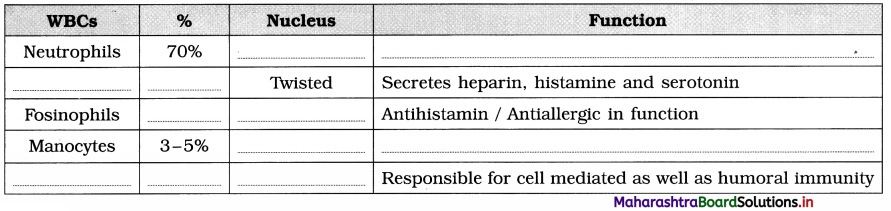
Answer:
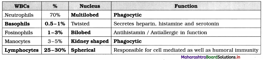
Question 4.
Complete the following flow chart
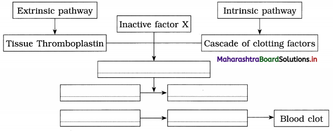
Answer:
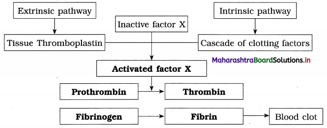
Question 5.
Complete the following Table
| Waves on ECG | Heart Activity | Caused due to |
| P wave | Atrial contraction | —————- |
| QRS wave | —————– | Ventricular depolarization |
| T wave | —————– | Ventricular repolarization |
Answer:
| Waves on ECG | Heart Activity | Caused due to |
| P wave | Atrial contraction | Atrial depolarization |
| QRS wave | Ventricular contraction | Ventricular depolarization |
| T wave | Ventricular contraction | Ventricular repolarization |
Question 6.
| Cardiovascular disorders | Symptom |
| Coronary Artery Diseases (CAD) | Deposition of calcium, fat, cholesterol and ——————— |
| —————- | Pain in chest resulting from reduction in the blood supply to the cardiac muscles. |
| Silent Heart Attack | Myocardial infarction without ———————- |
Answer:
| Cardiovascular disorders | Symptom |
| Coronary Artery Diseases (CAD) | Deposition of calcium, fat, cholesterol and fibrous tissues in blood vessels. |
| Angina pectoris | Pain in chest resulting from reduction in the blood supply to the cardiac muscles. |
| Silent Heart Attack | Myocardial infarction without showing symptoms of classical heart attack. |
![]()
Question 7.
| Instrument / Technique | Purpose of use |
| Sphygmomanometer | ——————— |
| —————- | X-ray imaging of the cardiac blood vessels to locate the position of blockages. |
| ————— | To measure ECG. |
Answer:
| Instrument / Technique | Purpose of use |
| Sphygmomanometer | To measure blood pressure. |
| Angiography | X-ray imaging of the cardiac blood vessels to locate the position of blockages. |
| Electrocardiograph | To measure ECG. |
Diagram based questions
Question 1.
Give the name and function of A and ‘B’ from the diagram given below
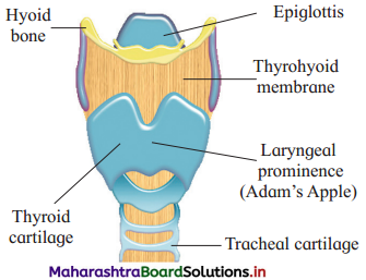
Answer:
| Name | Function |
| (A) Epiglottis | Epiglottis prevents the entry of food into trachea. |
| (B) Tracheal cartilage | Tracheal cartilage prevents collapse of trachea and always keeps it open. |
Question 2.
Label parts A’ and ‘B’ from the following diagram and answer the following questions
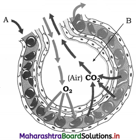
(a) What is the partial pressure of O2 in part ‘B’?
(b) What is the partial pressure of CO2 in part A’?
(c) How many alveoli are present in the lungs?
Answer:
Label A → Blood from pulmonary artery Label B → Alveolus
(a) The partial pressure of O2 in alveolar air is 104 mm Hg.
(b) The partial pressure of CO2 in pulmonary capillaries is 45 mmHg.
(c) There are about 700 million alveoli in the lungs.
Question 3.
Label parts A’ and ‘B’ from the given diagram and give their functions.
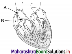
Answer: Part A → Sinoatrial [SA] node
Function : SA node acts as pacemaker of heart because it has the power of generating a new wave of contraction and making the pace of contraction.
Part B → Atrioventricular [AV] node
Function : Atrioventricular [AV] node acts as pace-setter of heart.
Question 4.
Sketch and label the dorsal (posterior) view of human heart.
Answer:
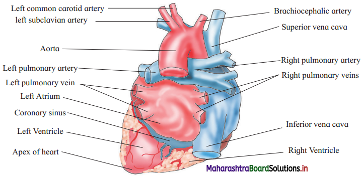
Question 5.
Sketch and label the ventral (anterior) view of human heart.
Answer:
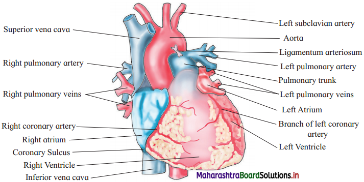
Question 6.
Sketch and label – Electrocardiogram or ECG.
Answer:
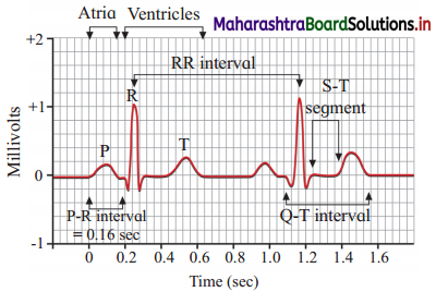
Question 7.
Sketch and label – T.S. of Artery, Vein and Capillary.
Answer:
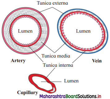
Question 8.
Observe the diagrams of blood cells and answer the following questions
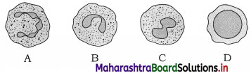
(a) Which of the above is agranulocyte ?
(b) Describe its origin and structure.
(c) Mention its types.
(d) Explain its function.
Answer:
(a) The figure ‘D’ is agranulocyte.
(b) Structure : Agranulocytes do not show cytoplasmic granules and their nucleus is not lobed.
(c) Agranulocytes are of two types, viz. lymphocytes and monocytes.
(d) Functions of agranulocytes : Agranulocytes are responsible for immune response of body by producing antibodies and monocytes are phagocytic in function.
Question 9.
Observe the diagrammatic representation of double circulation and answer the given questions.
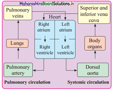
- Why the circulation shown in the above diagram is called double circulation?
- What are the two main routes of double circulation?
- Which blood vessels carry oxygented blood to heart and deoxygenated blood to lungs?
- Which blood vessels carry deoxygented blood to heart and oxygenated blood to body organs?
Answer:
- During circulation, blood passes twice through the heart, therefore it is called double circulation.
- (1) Pulmonary circulation which is from heart to lungs and back from lungs to heart.
(2) Systemic circulation which is from heart to body and back from all body organs to the heart. - Oxygenated blood is carried to the heart by pulmonary veins. Dexoygenated blood is carried to the lungs by pulmonary artery.
- Deoxygenated blood is carried to heart by superior and inferior vena cavae. Oxygenated blood is carried to the body organs by systemic or dorsal aorta.
![]()
Question 10.
Observe the cardiac cycle given below and answer the following questions
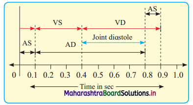
- Which phases of cardiac cycle are shown in the above diagrammatic representation ?
- How much time is taken for entire heart to be in diastole?
- How much longer is ventricular systole as compared to atrial systole?
Answer:
- There are four main phases of cardiac cycle shown in the above diagram. They are
(1) AS : Arterial systole. (2) AD : Atrial diastole. (3) VS : Ventricular systole.
(4) VD : Ventricular diastole which is along with joint diastole. - Diastole of entire heart is called joint diastole, which is for about 0.4 second.
- Ventricular systole is almost for the double time than the atrial systole. Atrial systole is for 0.15 second whereas ventricular systole is for 0.3 second.
Long Answer Questions
Question 1.
Describe the respiratory system of human.
Answer:
Respiratory system of human : Human respiratory system consists of nostrils, nasal chambers, pharynx, larynx, trachea, bronchi, bronchioles, lungs, diaphragm and intercostal muscles.
1. Nostrils and nasal chambers:
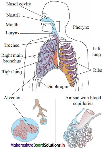
- Oxygen rich air is taken in the body through the nostrils or external nares. They are external opening of the nose. Carbon dioxide and water vapour are also released out of the body through the same passage i.e. the nostrils.
- Internal nares open into the pharynx. The space between external and internal nares is knows as nasal chamber which is lined internally by mucous membrane and ciliated epithelium.
- Nasal chamber is divided into two parts by a cartilage called mesethmoid. Each part of these halves is further divided into three regions, viz. vestibule, respiratory part and sensory part.
- Vestibule is the anteriormost part of nasal chamber. In the vestibule fine hairs are present. They filter out the dust particles and prevent them from going inside.
- Respiratory part is the second region which is richly supplied with the capillaries. Air is made warm and moist in this region.
- Sensory part is lined by sensory epithelium. It is concerned with the detection of smell.
2. Pharynx:
- Pharynx is a short and vertical tube measuring about 12 cm in length. In pharynx the respiratory and food passages cross each other.
- The upper part of pharynx is known as naso-pharynx which conducts the air. The lower part is called laryngo-pharynx or oro¬pharynx which conducts food to the oesophagus.
- Tonsils that are made up of lymphatic tissue are present in the pharynx. They kill the bacteria by trapping them in the mucus.
3. Larynx:
- Larynx produces sound. In males it increases in size at puberty. This is termed as Adam’s apple. It is clearly seen in the neck region.
- From pharynx air enters the larynx. The opening through which it enters is called glottis. Glottis is guarded by a flap called epiglottis.
- Epiglottis prevents the entry of food particles into the trachea.
- TWo folds of elastic tissue called vocal cords are seen along the side of glottis. When they vibrate the sound is produced.
4. Trachea:
- The trachea or wind pipe is about 12 cm long and 2.5 cm wide.
- It is situated in front of the oesophagus and runs downwards in the thorax through the neck.
- The trachea is made up of fibrous muscular tissue wall which is supported by ‘C’-shaped cartilages. These cartilaginous rings are 16 to 20 in number.
- Internally the tracheal wall bears ciliated epithelium and mucous glands.
- When any foreign particle enters the trachea inadvertently. It is thrown out by coughing action.
- Mucous and ciliary action remove the dust particles and push them upwards to the larynx. These particles are then gulped and taken into the oesophagus.
5. Bronchi and bronchioles:
- At the distal end, the trachea divides into two bronchi (Singular – bronchus). Bronchi lie below the sternum or breast bone.
- Each bronchus has a complete ring of cartilage for support. The two bronchi enter into the lungs on either side.
- After entering into the lungs each bronchus divides into secondary and tertiary bronchi. The tertiary bronchi divide and re-divide to form minute bronchioles.
- Bronchioles do not have cartilages in their walls. Each bronchiole ends into a balloon like alveolus.
- Owing to the presence of alveoli the lungs become spongy and elastic.
6. Lungs:
- Lungs are principal respiratory organs located in the thoracic cavity.
- They are pinkish, soft, hollow, paired, elastic and distensible organs.
- Each lung is enclosed in a pleural sac which consists of two membranes, viz. an outer parietal and inner visceral.
- The parietal and visceral membranes enclose pleural cavity which is filled with pleural fluid. The pleural fluid lubricates and prevents friction when pleural membranes slide on each other.
- Lungs are highly vascular as they are richly supplied with blood capillaries.
- The left lung has two lobes while the right lung has three lobes. Each lobe has many bronchioles and alveolar sacs. The alveolar sacs are spherical and thin walled.
- Each alveolar sac contains about 20 alveoli. The alveoli appear as a bunch of grapes. The lobule in the lung thus consists of alveolar ducts, alveolar sacs and alveoli.
- Each alveolus has thin and elastic walls. It is about 0.1 mm in diameter. Alveoli are covered by network of capillaries from pulmonary artery and pulmonary vein. A network of pulmonary capillaries supply the alveolus.
- The alveolar wall is 0.0001 mm thick and made up of simple, non-ciliated, squamous epithelium. It has collagen and elastin fibres.
- Every lung has about 700 million alveoli. They increase the surface area of the lungs for exchange of gases.
Question 2.
Describe the process of respiration in man.
OR
Describe the mechanism of respiration in human beings.
Answer:
Respiration includes breathing, external respiration, internal respiration and cellular respiration.
A. Breathing : During breathing air comes in and goes out of the lungs. The rate of gaseous exchange is speeded up by breathing. Breathing involves two processes, viz. inspiration and expiration
1. Inspiration:
- Inspiration is the process in which the air containing oxygen is taken inside the lung.
- Inspiration is the active process which is possible due to intercostal muscles, sternum and diaphragm.
- During inspiration, intercostal muscles contract, ribs are pulled outward as a result of which the space in the thoracic cavity is increased.
- At the same time the lower part of the breast bone is raised and diaphragm flattens by contraction.
- The volume of thoracic cavity is thus increased.
- Pressure in the lungs decreases as the lungs expand and their volume is increased. Owing to this the atmospheric air enters inside the body through respiratory passage and reaches the lungs.
2. Expiration:
- Expiration is the process in which air containing carbon dioxide and water vapour is expelled out of the lungs.
- Expiration is a passive process.
- During expiration, intercostal muscles relax and ribs are pulled inwards.
- The diaphragm relaxes and becomes dome shaped. Intercostal muscles contract simultaneously and due to these events, the volume of the thoracic cavity is reduced.
- The pressure on the lungs is thus increased as a result of which they are compressed.
- Due to this, air rushes out of the lungs and is expelled out through the nose.
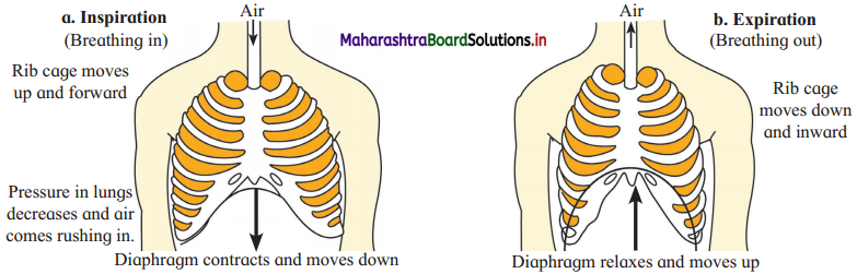
B. External respiration : The process of external respiration takes place in the lungs where oxygen from the lungs diffuses into the blood capillaries present in the lung tissue. Similarly carbon dioxide from the blood capillaries diffuses out and enters in the alveoli in the lungs.
C. Internal respiration : Internal respiration takes place in the cells of the body. Oxygen brought by the blood is given to the cells and the tissues during internal respiration. Similarly carbon dioxide passes into the blood cells from the cells and tissues.
D. Cellular respiration : Cellular respiration takes place in the mitochondria of the cell, where oxygen is utilised to liberate energy in the form of ATP molecules.
Question 3.
Describe the respiratory disorders.
Answer:
- Following are some respiratory disorders : Emphysema is caused due to alveolar abnormalities. Chronic bronchitis results into coughing and shortness of breath.
- Viral and bacterial respiratory diseases. Acute bronchitis, sinusitis, laryngitis and pneumonia are some of the inflammatory diseases caused either due to virus or due to bacteria.
- Allergens like pollen or pet dander can cause asthma. In asthma constriction of bronchioles takes place causing periodic wheezing and difficulty in breathing.
- Occupational hazards cause respiratory diseases like silicosis or asbestosis. In these disorders there is inflammation fibrosis leading to lung damage.
Treatments of respiratory diseases:
- Bacterial diseases can be completely cured by specific antibiotics.
- Viral diseases need to be taken care of by using vaporizers and decongestants.
- Asthma needs treatment by inhalers and nebulizers.
- For occupational disorders proper mask and other protective gear is a must.
- Lethal diseases like pneumonia should be controlled by medication and rest.
Question 4.
How does transport of O2 and CO2 take place in man?
Answer:
1. Transport of O2:
- Only 3% of the total oxygen is transported in a dissolved state by the plasma.
- The remaining 97% is transported in the form of oxyhaemoglobin in the RBCs.
- Hemoglobin present is RBCs combines with oxygen to form oxyhaemoglobin.
Hb + 4O2 → Hb (4O2) - Oxyhaemoglobin is transported from lungs to the tissues where it readily dissociates to release O2.
- Binding of oxygen with haemoglobin in the alveoli and release of oxygen into the tissue cells depends upon the difference in partial pressure of O2 and CO2.
2. Transport of CO2: Carbon dioxide is transported by RBCs and plasma in three different forms.
- By plasma in solution form (7%) : About 7% of CO2 is transported in a dissolved form as carbonic acid (which can be broken down into CO2 and H2O).
CO2 + H2O = H2CO3. - By bicarbonate ions (70%) : Nearly 70% of carbon dioxide is transported in the form of sodium bicarbonate/potassium bicarbonate in the plasma.
- RBCs contains an enzyme, carbonic anhydrase. In the presence of this enzyme CO2 combines with water to form carbonic acid.
- Carbonic anhydrase also brings about dissociation of carbonic acid immediately tending to large accumulation of HCO3- ions inside the RBCs.
CO2 + H2O Carbonic anhydrese H2CO2 Carbonic anhydrase H+ + HCO3- - The bicarbonate ions moves out of RBCs and this would bring about imbalance of the charge inside the RBCs.
- To maintain the ionic balance, Cl– ions diffuse from plasma into the RBCs. This movement of chloride ions is known as chloride shift or Hamburger’s phenomenon.
- HCO3- ions from the plasma then joins to Na+/K+ forming NaHCO3/KHCO3 (to maintain PH of blood).
HCO3- + Na+ → NaHCO3 Sodium bicarbonate - H+ is taken up by haemoglobin to form Reduced Hb (HHb).
- At the level of the lungs due to the low partial pressure of the alveolar air, hydrogen ion and bicarbonate ions recombine to form carbonic acid and in presence of carbonic anhydrase it again yields carbon dioxide and water.
H+ + HCO3- Carbonic anhydrase H2CO3 Carbonic anhydrase CO2 + H2O.
3. By red blood cells (23%):
- Carbon dioxide binds with the amino group of the haemoglobin and form a loosely bound compound carbaminohaemoglobin Hb + CO2 = HbCO2
- Due to low partial pressure of CO2 at alveolus carbaminohaemoglobin decomposes releasing the carbon dioxide.
![]()
Question 5.
Describe different types of leucocytes.
OR
Describe five types of leucocytes, with the help of diagrams. Add a note on their functions.
Answer:
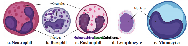
- Leucocytes or White Blood Corpuscles (WBCs) are colourless, nucleated, amoeboid and phagocytic cells.
- Their size ranges between 8 to 15 pm. Total WBC count is 5000 to 9000 WBCs/cu mm of blood. The average life span of a WBC is about 3 to 4 days.
- They are formed by leucopoiesis in red bone marrow, spleen, lymph nodes, tonsils, thymus and Payer’s patches, whereas the dead WBCs are destroyed by phagocytosis in blood, liver and lymph nodes.
- Leucocytes are mainly divided into two types, viz., granulocytes and agranulocytes.
- Granulocytes : Granulocytes are cells with granular cytoplasm and lobed nucleus. Based on their staining properties and shape of nucleus, they are of three types, viz. neutrophils, eosinophils and basophils.
(I) Neutrophils:
- In neutrophils, the cytoplasmic granules take up neutral stains.
- Their nucleus is three to five lobed.
- It may undergo changes in structure hence they are called polymorphonuclear leucocytes or polymorphs.
- Neutrophils are about 70% of total WBCs.
- They are phagocytic in function and engulf microorganisms.
(II) Eosinophils or acidophils:
- Cytoplasmic granules of eosinophils take up acidic dyes such as eosin. They have bilobed nucleus.
- Eosinophils are about 3% of total WBCs.
- They are non-phagocytic in nature.
- Their number increases (i.e. eosinophilia) during allergic conditions.
- They have antihistamine property.
(III) Basophils:
- The cytoplasmic granules of basophils take up basic stains such as methylene blue.
- They have twisted nucleus.
- In size, they are smallest and constitute about 0.5% of total WBCs.
- They too are non-phagocytic.
- Their function is to release heparin which acts as an anticoagulant and histamine that is involved in inflammatory and allergic reaction.
Agranulocytes : There are two types of agranulocytes, viz. monocytes and lymphocytes. Agranulocytes do not show cytoplasmic granules and their nucleus is not lobed. They are of two types, viz. lymphocytes and monocytes.
(I) Lymphocytes:
- Agranulocytes with a large round nucleus are called lymphocyte.
- They are about 30% of total WBCs.
- Agranulocytes are responsible for immune response of the body by producing antibodies.
(II) Monocytes:
- Largest of all WBCs having large kidney shaped nucleus are monocytes. They are about 5% of total WBCs.
- They are phagocytic in function.
- They can differentiate into macrophages for engulfing microorganisms and removing cell debris. Hence they are also called scavengers.
- At the site of infections they are seen in more enlarged form.
Question 6.
Give an account of external features of the human heart.
Answer:
(1) The heart is hollow, muscular, conical organ about the size of one’s fist with broad base and narrow apex tilted towards left measuring about 12 cm in length. 9 cm in breadth and weighing about 250 to 300 grams.
(2) The human heart has four chambers, two atria which are superior, small, thin walled receiving chambers and two ventricles which are inferior, large, thick walled, distributing chambers.
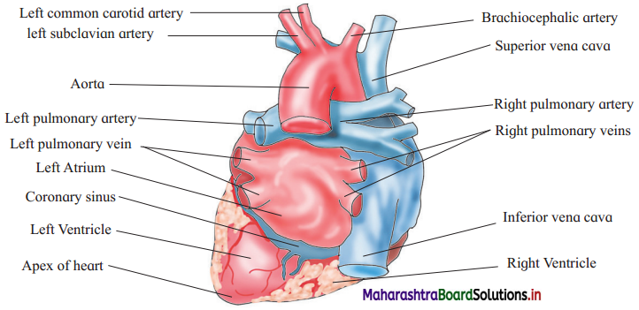
(3) Externally there is a transverse groove between the atria and the ventricles which is known as atrioventricular groove or coronary sulcus.
(4) Between the right and left ventricles there is interventricular sulcus (pi. sulci). In these sulci the coronary arteries and coronary veins are present.
(5) Oxygenated blood to the heart is supplied by coronary arteries while coronary veins collect deoxygenated blood from the heart. The coronary veins join to form coronary sinus which opens into the right atrium.
(6) Right atrium is larger in size than the left atrium. Deoxygenated blood from all over the body is brought through superior vena cava and inferior vena cava and poured into right atrium. Oxygenated blood from lungs is brought to heart by two pairs of pulmonary veins which carry it to the left atrium.
(7) Pulmonary trunk is seen arising from the right ventricle, which carries deoxygenated blood to lungs. While systemic aorta arises from the left ventricle and carries oxygenated blood to all parts of the body.
(8) The pulmonary trunk and systemic aorta are connected by ligamentum arteriosum that represents remnant of ductus arteriosus of foetus.
Question 7.
With the help of well labelled diagram describe the internal structure of human heart.
OR
Sketch and label internal view of heart.
Answer:
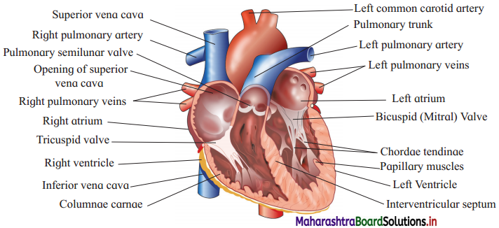
The heart shows four chambers with two atria and two ventricles.
I. Atria:
1. Right atrium:
- There are two atria which are separated from each other by interatrial septum. They are thin walled receiving chambers on the upper side.
- The right atrium receives deoxygenated blood from upper part of body through superior vena cava and from the lower part of the body by inferior vena cava. In the right atrium opens the coronary sinus.
- Eustachian valve guards the opening of inferior vena cava while opening of coronary sinus is guarded by Thebesian valve.
- On the right side of interatrial septum is seen an oval depression called the fossa ovalis. In the interatrial septum of the foetus there is an oval opening called foramen ovale. Fossa ovalis is remnant of this foramen ovale.
- Right atrium opens into the right ventricle.
2. Left atrium:
- The oxygenated blood from the lungs is brought into left atrium through four openings of pulmonary veins.
- Left atrium opens into the left ventricle.
II. Ventricles:
- There are two ventricles which are separated from each other by interventricular septum. They are two thick walled distributing chambers situated on the lower side of the heart.
- Left ventricle has thickest wall as it pumps blood to all parts of the body.
- The inner surface of the ventricle is thrown into a series of irregular muscular ridges called columnae carnae or trabeculae carnae.
- Each atrium opens into the ventricle of its side through atrioventricular aperture. These apertures are guarded by valves made up of connective tissue. The right atrioventricular valve has three flaps hence called tricuspid valve. Left atrioventricular valve has two flaps hence called bicuspid valve or mitral valve.
- Bicuspid and tricuspid valves are attached to papillary muscles of ventricles by chordae tendinae. The chordae tendinae prevent the valves from turning back into the atria during the contraction of ventricles.
- From the right ventricle arises pulmonary trunk which carries deoxygenated blood to lungs for oxygenation.
- From the left ventricle arise systemic aorta which distributes oxygenated blood to all parts of the body.
- Pulmonary aorta and systemic aorta has three semilunar valves at the base which prevent backward flow of blood during ventricular diastole.
Question 8.
With the help of suitable diagram, describe the conducting system of human heart.
Answer:
(1) The human heart is myogenic.
(2) Conducting system of the heart consists of sinoatrial node, atrioventricular node, bundles of His and Purkinje’s fibre system.
(3) The pacemaker of the heart is sinoatrial node because here the heartbeat originates. Pacemaker has power of generation of wave of contraction. This is modified cardiac tissue, also called a nodal tissue.
(4) SA node is situated in the wall of right atrium near the opening of superior vena cava. The wave of contraction generated by SA node is conducted by cardiac muscle fibres to both the atria. This results in contraction resulting into atrial systole.
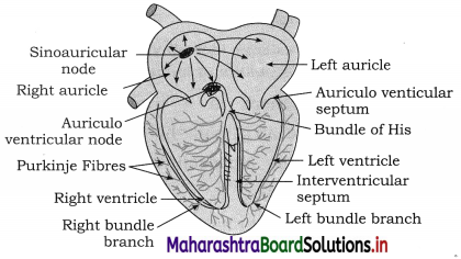
(5) The atrioventricular node (AV node) is located in the wall of right atrium near the opening of coronary sinus. AV node receives the wave of contraction generated by SA node through intermodal pathways.
(6) Bundle of His arises from AV node and divides into right and left bundle branches. These are located in the interventricular septum.
(7) The bundle branches further form Purkinje fibres which penetrate into myocardium of ventricles.
(8) The bundle of His and Purkinje fibres conduct the wave of contraction from AV node to myocardium of ventricles causing ventricular systole.
![]()
Question 9.
Describe the detail cardiac cycle.
OR
Explain the working of heart.
Answer:
(1) The working of heart or cardiac cycle is formed by atrial systole, ventricular systole and joint diastole. It takes place in 0.8 second.
(2) During atrial systole from right atrium, the deoxygenated blood is poured into right ventricle through atrioventricular aperture. Similarly, from left atrium, the oxygenated blood enters the left ventricle through atrioventricular aperture. This entire atrial systole lasts for 0.1 second.
(3) After auricular systole follows the ventricular systole. During ventricular systole, the deoxygenated blood from the right ventricle enters the pulmonary trunk, which carries blood to lungs for oxygenation. At the same time, the oxygenated blood from the left ventricle enters the aorta which is then supplied to all parts of the body. The ventricular systole lasts for 0.3 second.
(4) Joint diastole or complete cardiac diastole is the phase taking place after the systole, when the entire heart undergoes relaxation, for 0.4 second.
(5) During joint diastole, the right atrium receives deoxygenated blood from all parts of the body through superior vena cava, inferior vena cava and coronary sinus. The left atrium receives oxygenated blood from the lungs through two pairs of pulmonary veins.
Question 10.
What is blood clotting? How and when does it occur?
Answer:
- Blood clotting is coagulation of blood in order to stop the blood flow and resuting blood loss at the time of injury.
- When the blood vessel is intact, blood does not clot due to the presence of active anticoagulants like heparin and antithrombin. But when there is an injury causing rupture of a blood vessel, bleeding starts.
- This bleeding is stopped by the process of blood clotting during which liquid blood is converted into semisolid jelly.
The events occurring during blood clotting are as follows:
- Release of thromboplastin from thrombocytes and injured tissue.
- Formation of enzyme prothrombinase in the blood due to initiation of thromboplastin.
- Conversion of inactive prothrombin into active thrombin by prothrombinase in the presence of Ca ions.
- Conversion of soluble fibrinogen into insoluble fibrin by thrombin.
- Formation of a clot by enmeshing platelets, other blood cells and plasma in the fibrin fibres enmesh.
These reactions occur in 2 to 8 minutes. Therefore, clotting time is said to be 2 to 8 minutes.
Question 11.
What is repolarization and depolarization ?
Answer:
Repolarization is a stage of an action potential in which the cell experiences a reduction of voltage due to the efflux of potassium (K+) ions along its electrochemical gradient. This phase occurs after the cell reaches its highest voltage from depolarization.
Depolarization occurs in the four chambers of the heart : both atria first and then both ventricles. The SA node sends the depolarization wave to the atrioventricular (AV) node which-with about a 100 minutes delay to let the atria finish contracting-then causes contraction in both ventricles, seen in the QRS wave.
Question 12.
What is the correlation between depolarization and repolarization as well as contraction and relaxation of the heart?
Answer:
Depolarization and Repolarization:
- When cardiac cells are at rest, they are polarized, meaning no electrical activity takes place.
- The cell membrane of the cardiac muscle cell separates different concentrations of ions, such as sodium, potassium, and calcium. This is called the resting potential.
- Electrical impulses are generated by specialized cardiac cells automatically.
- Once an electrical cell generates an electrical impulse, this electrical impulse causes the ions to cross the cell membrane and causes the action potential, also called depolarization.
- The movement of ions across the cell membrane through sodium, potassium and calcium channels, is the drive that causes contraction of the cardiac cells/muscle.
- Depolarization with corresponding contraction of myocardial muscle moves as a wave through the heart. Depolarization thus corresponds with contraction of heart.
- Repolarization is the return of the ions to their previous resting state, which corresponds with relaxation of the myocardial muscle. Repolarization thus corresponds with relaxation of heart.
- Depolarization and repolarization are electrical activities which cause muscular activity.
- The electrical changes in the myocardial cell during the depolarization – repolarization cycle is detected on ECG.
![]()
Question 13.
How are the signals detected and amplified by electrocardiograph?
Answer:
- The action potential created by contractions of the heart wall spreads electrical currents from the heart throughout the body.
- The spreading electrical currents create different potentials at points in the body, which can be sensed by electrodes placed on the skin.
- The electrodes are made of metals and salts and they act as biological transducers.
- Ten electrodes are attached to different points on the body while taking ECG.
- There Eire three main leads responsible for measuring the electrical potential difference between arms and legs.
- Electrical potential difference between electrodes is recorded.
- As in all ECG lead measurements, the electrode connected to the right leg is considered the ground node.
- These ECG signals are acquired using a biopotential amplifier and then displayed using instrumentation software. This is recorded on ECG machine or electrocardiograph. The recorded ECG is anailysed by an expert.